Download
1 / 29
300 likes | 460 Views
Psychology 4051. Visual Functions. Stereopsis. True depth perception. The ability to see in 3D. Does not include monocular cues or binocular kinetic cues. Interposition. Relative Size. Linear Perspective. Stereopsis. Occurs due to the differing positions of the eyes.

E N D
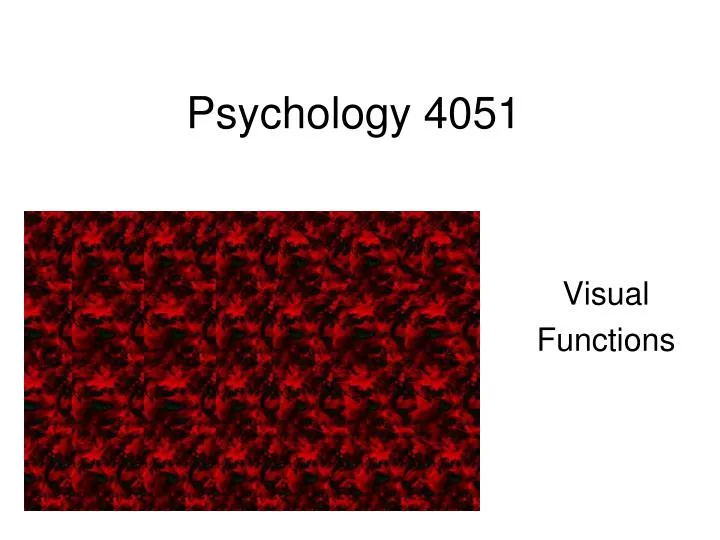
Psychology 4051 Visual Functions
Stereopsis • True depth perception. The ability to see in 3D. • Does not include monocular cues or binocular kinetic cues. Interposition Relative Size Linear Perspective
Stereopsis • Occurs due to the differing positions of the eyes. • Retinal disparity: An image will fall on noncorresponding retinal areas.
Stereopsis • Horopter: an imaginary arc upon which object produce no disparity. • Objects along the horopter do not produce disparity. • Those in front produce crossed disparity. • Those in back produce uncrossed disparity.
Stereopsis • Panum’s Fusional Area: Images from object within the area can be fused. • Those outside the area produce diplopia.
Stereopsis • Fusion is accomplished by disparity selective cells. • They respond to specific amounts of disparity and produce the sensation of depth.
Measurement of Stereopsis • Stereoacuity: the minimum amount of disparity one can use to detect depth. • Measured in seconds of arc (arc sec). • Adult stereoacuity is less than 40 arc sec. • Measured with random dot stereograms. Arrays of dots that appears to have a patternless texture.
Measurement of Stereopsis • A portion of the stereogram contains two patterns that are displaced laterally. • When viewed with polarized glasses, the lateral displacement creates artificial retinal disparity. • i.e., a slightly different image is seen by each eye. • Creates the sensation of depth.
Measurement in Young Children • Measurement in preschoolers can be conducted using the Randot Preschool Stereoacuity Test. • Consists of three books that contain random dot stereograms. • Each stereogram is in the form of a shape that is familiar to children. • Hands, hearts, ducks, elephants, etc. • Shapes cover a broad range of retinal disparities • 800 to 40 arc sec
Measurement in Young Children • Infants and toddlers can be tested with Randot Stereo Smile Cards. • Modeled on the Teller Acuity Cards • The cards are completely covered with a random dot array. • When viewed through polarized glasses, one side of the card possesses a happy face of crossed disparity. • 480 to 120 arc sec
Measurement in Young Children • The cards are presented following FPL. • Very difficult to measure stereoacuity in infants and toddlers. • The target is not very salient.
Development • Stereopsis emerges at 3.5 to 6 months of age and shows rapid improvement. • Then rises slowly to adult levels. • Measures 100 arc sec at 3 years, 50 arc sec at 5 years, and 40 arc sec at 7 years.
Development • May be correlated with the segregation of ocular dominance columns. • At birth, neurons from both eye converge onto single neurons in layer IV in the visual cortex. • Later, there is a segregation of these connections, and connections separate into left eye and right eye columns. • i.e., ocular dominance columns.
Development • Convergence of connections occurs at the next level. • Animal studies show that stereopsis occurs at the same time this segregation into ocular dominance columns occurs.
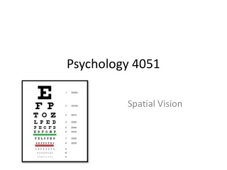
Refractive Error • The degree of myopia, hyperopia, or astigmatism. • Measured in diopters. • The refractive power of a lens. • The reciprocal of focal length. • The focal length of the eye is approximately 17 mm.
Refractive Error • The required refractive power of the eye is 60 D.
Refractive Error • Emmetropia: No refractive error. • Ametropia: Presence of refractive error. Spherical Refractive Error: Due to a mismatch between the focusing power of the eye and the length of the eye. • Reported relative to the 60 D norm.
Refractive Power • Myopia: the eye is too long to match its focal power. The image is focused in front of the retina. • Corrected with a concave lens.
Refractive Error • Focal length is longer than 17 mm • Required refractive power will be less than 60 D. • E.g. 57 D. • Corrected with a concave lens with – 3D power.
Refractive Power • Hyperyopia: the eye is too short to match its focal power. The image is focused behind the retina. • Corrected with a convex lens.
Refractive Error • Focal length is shorter than 17 mm • Required refractive power will more than 60 D. • E.g. 63 D. • Corrected with a convex lens with + 3 D power.
Refractive Error • Cylindrical Refractive Error • Astigmatism: a distortion in the shape of the cornea. • The cornea is curved more sharply along one axis than along the other. • As a result, the image is distorted. • Can be corrected by using a lens that counteracts the distortion
Measuring Refractive Error • Cycloplegic Retinoscopy: Beam of light is shone through the subject’s optical system. • Ophthalmologist looks through the site hole and observes the reflected light and a shadow. • A mirror inside the retinoscope is moved in various direction. • Movement of the shadow is observed
Measurement in Infants and Toddlers • Usually conducted using cycloplegia which prevents accommodation. • Can be difficult in infants and toddlers. • Can be conducted without cycloplegia, but requires expertise. • They can be assessed using a photoscreener (photorefractor).
Measurement in Infants and Toddlers • Camera and a flash source that take a photograph of the flashed light as it return from its passage through the optical system. • Based on the position and amount of crescent shaped light reflected from the subject’s pupil, refractive error can be determined.
Measurement in Infants and Toddlers • Can also be measured using an automatic refractor or autorefractor. • An infrared beam is shone into the eye. • The reflected beams return to the autorefractor and determines the extent to which the beam is out of focus.
Measurement in Infants and Toddlers • Both techniques are fast, objective, and require little expertise. • Neither is as accurate as cycloplegic retinoscopy.
Development • We are born farsighted (hyperopic). • This error is reduced through emmetropization as the eye grows. • 1 month = 2.2 D (Mayer et al., 2001) • 1 year = 1.57 D • 2 years = 1.19 D • 3 years = 1.00 D • 4 years = 1.13 D
Development • The length of the eye increases rapidly at first during the "infantile" high-growth period and then more slowly during the "juvenile" slow-elongation period. • This moves the retina away from the cornea so that, eventually, the length matches the focal power, producing emmetropia. • The growth of the eye appears to be controlled by the amount of blur of the image it receives.