Download
1 / 32
340 likes | 694 Views
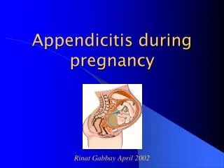
Sadaf Alipour General Surgeon Assistant Professor Tehran University of Medical Sciences. Appendicitis in pregnancy. The most common general surgical problem during pregnancy Incidence %0.06- 0.1 percent, or 1 in 1500 deliveries

E N D
Sadaf Alipour General Surgeon Assistant Professor Tehran University of Medical Sciences Appendicitis in pregnancy
The most common general surgical problem during pregnancy Incidence %0.06- 0.1 percent, or 1 in 1500 deliveries However, less in pregnant women than in age-matched nonpregnants Slightly higher rate in T2 than T1, T3 or postpartum More likely to rupture, especially in T3, possibly because of delay in Dx and intervention INTRODUCTION
Similar to nonpregnants RLQ pain: the most common symptom Should alert the physician caring for the pregnant to strongly consider apx Pain is close to McBurney's point in most regardless of stage of Py although appendix migrates a few cm cephalad with the enlarging uterus CLINICAL MANIFESTATIONS(1)
NL:Abdominal discomfort in Py due to enlarging uterus, fetal position or movement, Braxton-Hicks Severe, sudden, constant pain with other symptoms (nausea, vomiting, vaginal bleeding) or in upper abdomen suggests a disease. Peritoneal signs (rebound , guarding) never NL in Py Nausea and vomiting: common in early Py, usu abate by early to middle T2 but not normal when with abdominal pain, fever, diarrhea, headache, or localized abdominal findings CLINICAL MANIFESTATIONS(2)
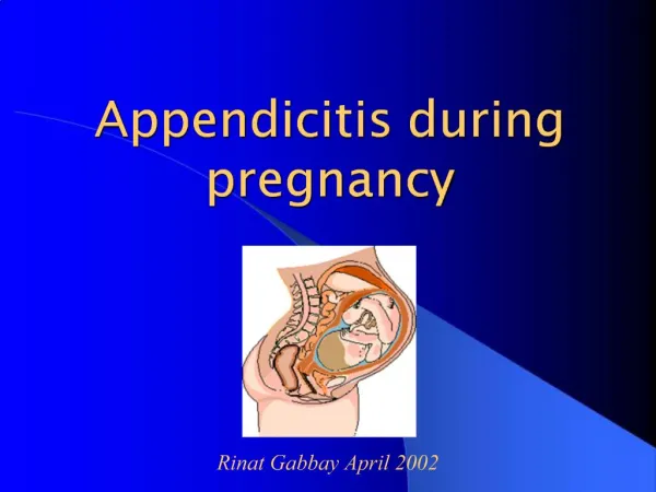
Physiologic changes of Py may affect presentation Uterus becomes abdominal, enlarging beyond pelvis by 12 weeks Uterus impedes examination and affect NL location of pelvic and abdominal organs CLINICAL MANIFESTATIONS(3)
Gravid uterus lifts anterior abdominal wall • Less direct contact between area of inflammation and parietal peritoneum • Less muscle response or guarding • Less peritoneal findings than nonpregnants • The laxity of the abdominal wall may also diminish peritoneal signs. CLINICAL MANIFESTATIONS (4)
Normal Py in T1 and T2 : WBC =6000- 16,000 , may rise to 20,000 -30,000 during labor Leukocytosis may NL in Py but bandemia not NL in Py and suggests infection until proven otherwise Retrospective review of 66,993 deliveries with 67 with probable Dx of apx: in those with confirmed apx, mean WBC=16,400 -versus 14,000 for those without apx. Laboratory assessment(1)
Inflamed appendix often close to bladder and ureter Microscopic hematuria and pyuria in up to one-third of acute appendicitis Pregnants with pyuria may be treated for UTI and forgo further investigation, delaying Dx of apx Laboratory assessment(2)
Especially difficult, requires high index of suspicion. Labor can be associated with pain that may be lateralized, May fever, leukocytosis, and vomiting when chorioamnionitis during labor diagnosis of apX in a laboring patient
IfDx unclear after assessment of complaints, examination, and lab: diagnostic imaging necessary as in nonpregnants Thus, virtually all pregnant women will have an imaging study Imaging
Choice for imaging of appendix in Py: graded compression ultrasonography Allows visualization of uterus, placenta and ovarie Can exclude other causes of RLQ pain Apx diagnosed if noncompressible blind- ended tubular structure in RLQ with diameter greater than 6 mm . As a general rule, if a normal appendix is not visualized, appendicitis cannot be excluded Ultrasonography
overall sensitivity=%86 Specificity=%81 However, gravid uterus can interfere with US, esp in the T3, leading to high negative laparotomy rate when US results inconclusive In one small series, appendix could not be visualized with US in 22 of 23 pregnants with suspected apx US
Where available, useful for the next step in diagnostic uncertainty MRI is an alternative to CT because it avoids exposure to ionizing radiation. Observational data suggest that MRI can accurately diagnose appendicitis during pregnancy MRI (1)
Excellent modality for excluding apx in Py with characteristic signs and symptoms when inconclusive US Gadolinium not routinely administered because of theoretical fetal safety concerns, but may be used if essential . If a prolonged wait before MRI, increasing risk of rupture over time should be considered and undue delays for imaging avoided. MRI (2)
Sensitivity= %100 Specificity= %93 Positive predictive value = %61 Negative predictive value = %100 MRI (3)
Main findings of apx on CT: • RLQ inflammation • Enlarged nonfilling tubular structure • Appendicolith. CT scan (1)
Modifications of CT protocol can limit fetal exposure to less than 3 mGy (30 mGy for carcinogenesis in fetus) Standard abdominal CT with oral and IV contrast or a specialized appendiceal CT protocol can also be used, but are associated with higher fetal radiation exposure (20 to 40 mGy) CT scan (2)
Overall sensitivity= %94 Specificity= % 95 We suggest CT when clinic and US are inconclusive and MRI is not available Ct scan (3)
Decision for laparotomy should be based on clinic, imaging results, and clinical judgment Lab not particularly useful ecxept for R/O of alternate diagnoses Delaying Sx for more than 24 h increases risk of perforation (%14-43 of such patients) MANAGEMENT APPROACH AND OUTCOME
When Dx relatively certain: transverse incision at McBurney's point, or more commonly, over point of maximal tenderness When Dx less certain: lower midline vertical incision incision
Several case reports and small case series: laparoscopic appendectomy in Py feasible in all trimesters and with few complications One systematic review: higher rate of fetal loss with laparoscopy than open appendectomy, but data were from retrospective series Laparoscopy(1)
Decision to proceed to laparoscopy based on: • skill and experience of surgeon • clinical factors such as size of gravid uterus. Laparoscopy(2)
Risk of fetal loss higher in perforated apx (%36 versus %1.5)or when generalized peritonitis or abscess(fetal loss:% 6 versus %2; early delivery: %11 versus %4). Given diagnostic difficulties and significant risk of fetal mortality with perforation, a higher negative laparotomy rate (20 to 35 percent) compared to nonpregnant women has generally been considered to be acceptable. complications (1)
Maternal morbidity low except in perforated apx • Py related complications frequent in T1 and T2 • Spontaneous abortion %33 percent in T1 • Premature delivery %14 in T2 • No pregnancy complications in T3 complications (2)
C/S rarely indicated at time of appendectomy Risk of dehiscence during labor and vaginal delivery not increased when fascia appropriately reapproximated Type of delivery
Good long-term prognosis No increased risk of infertility or other complications prognosis
Free perforation causes intraperitoneal dissemination of pus and fecal material Patients quite ill and may be septic Increased risk of preterm labor and delivery and fetal loss Urgent laparotomy necessary with appendectomy and irrigation and drainage of the peritoneal cavity Perforated appendix
When contained perforation: treated with ABs , IV fluids, bowel rest, and close monitoring Many will respond since it has already been "walled-off.“ Although there is good evidence to support this approach in nonpregnant individuals, there is only limited evidence in pregnant women. In Nonpregnants with long duration of symptoms (more than five days)
Report of 2 patients: ABs (ampi, genta,clinda),IV fluids, and bowel rest: improvement of symptoms over 2-3 d In one: interval apy 2 m after NVD In the other: apy at c/s (breech with preterm labor In both: avoidance of glucocorticoids and tocolytics due to concerns of suppressing manifestations of worsening infection and delaying delivery if intraamniotic infection was also present. Until further experience, these should be followed closely in hospital to monitor for maternal sepsis and preterm labor. Conservative tx of apx in Py
Apx: most common general Sx problem in Py, clinic and Dx similar to nonpregnant RLQ pain within a few cm of McBurney's : most common symptom Nausea/vomiting: both apx and NL Py. In apx, following pain, in Py usu no pain. US: the best - noncompressible 6mm or more blind ended tubular structure in RLQ If clinic and US inconclusive: MRI, When MRI not available: CT Decision to proceed to Sx based on imaging and clinical judgment. Lab not particularly useful other than R/O other diagnoses. Delaying Sx more than 24 hours increases risk of perforation. When Dx relatively certain:transverse incision over point of maximal tenderness . When less certain: lower midline vertical incision SUMMARY AND RECOMMENDATIONS
1- Schwartz Principles of Surgery (book) 2-UptoDate (online) references