Download
1 / 27
320 likes | 446 Views
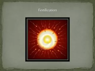
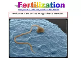
FERTILIZATION. After ovulation, the ovum, which is about 0.15 mm in diameter, passes into the uterine tube and is moved along towards the uterus. At sexual intercourse about 300 million sperms are deposited in the posterior fornix of the vagina

E N D
After ovulation, the ovum, which is about 0.15 mm in diameter, passes into the uterine tube and is moved along towards the uterus
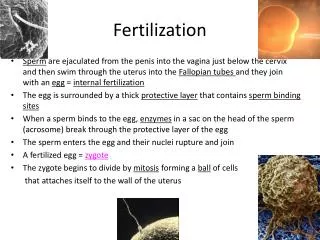
At sexual intercourse about 300 million sperms are deposited in the posterior fornix of the vagina • Some of the sperms survive & propel towards the uterine tube while the remaining sperms are destroyed by the acid medium of the vagina • More will die on the journey through the uterus, and only thousand sperms reach the uterine tube, where they meet the ovum, usually in the ampulla
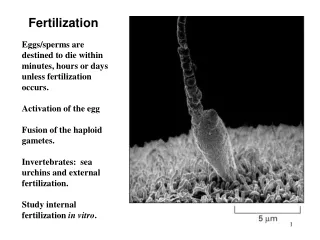
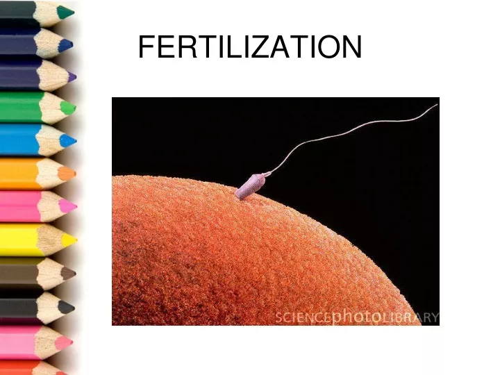
During this journey the sperms will become mature and release the enzyme hyaluronidase, which allows the penetration of the zona pellucida and the cell membranes surrounding the ovum
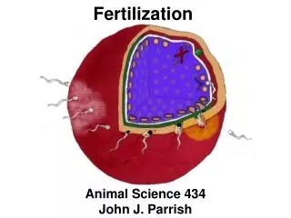
Many sperms are needed for this to take place, but only one will enter the ovum
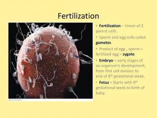
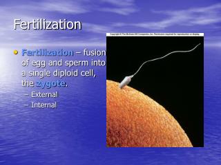
After this, the membrane is sealed to prevent entry of any further sperm and the nuclei of the two cells fuse • Sperm and the ovum each contribute half the complement of chromosomes to make a total of 46 • The sperm and ovum are known as the male and the female gametes and the fertilized ovum as the zygote
When the ovum has been fertilized ,it continues its passage through the uterine tube and reaches the uterus 3 or 4 days later • During this time cell division or segmentation takes place • Fertilized ovum divides into 2 cells, then into 4 , then 8 & 16 and so on until a cluster of cells is formed known as the morula ( mulberry )
Next a fluid filled cavity or blastocele, appears in the morula, which is known as blastocyst
Around and outside the blastocyst ,there is a single layer of cells known as the trophoblast • The remaining cells are clumped together at one end forming the inner cell mass
The trophoblast will become sticky and adheres to the endometrium • It begins to secrete substances that digest the endometrial cells , allowing the blastocyst to become embedded in the endometrium. • Embedding / nidation is normally complete by 11th day
Decidua • Decidua is the name given to endometrium during pregnancy • From, the time of conception , the increased secretions of oestrogens causes the endometrium to grow to 4 times its non – pregnant thickness
Progesterone stimulates the secretory activity of the endometrial glands and increase the size of the blood vessels • This accounts for the soft, vascular , spongy bed in which the fertilized ovum implants • Three layers are formed
The basal layer • Lies above the myometrium The functional layer • Layer consists of glands which are rich in secretions • Provides a secure anchorage for the placenta The compact layer • Layer forms the surface of the decidua
Trophoblasts • Small projections all over the surface of the blastocyst 3 Layers The syncytiotrophoblast • Layer composed of nucleated protoplasm , which is capable of breaking down tissues as in the process of embedding • Erodes the walls of the blood vessels of the decidua. Making the nutrients in the maternal blood available to the foetus
The cytotrophoblast • Produces hormone human chorionic gonadotrophin ( HCG ) • HCG informs corpus luteum that a pregnancy has begun • Continues to produce oestrogen and progesterone
Progesterone maintains the integrity of the decidua ,so that shedding does not takes place • i.e. menstruation is suppressed • High level of oestrogen suppresses production of FSH
The mesoderm • Layer consists of loose connective tissues
Inner cell mass form the foetus • 3 layers The ectoderm • Forms the skin and the nervous system The Mesoderm • This layer forms bones and muscles and also the heart and the blood vessels • certain internal organs also originate The Endoderm • Forms mucous membrane and glands
The 3 types together known as the embryonic plate • Two cavities appear in the inner cell mass : one on either side of the embryonic plate The amniotic cavity • Lies on the side of the ectoderm • Filled with fluid and gradually enlarges and folds around the embryo to enclose it
The yolk sac • lies on the side of the endoderm • Provides nourishment for the embryo ( Developing off spring after implantation and until 8 weeks after conception)