Download
1 / 23
240 likes | 257 Views
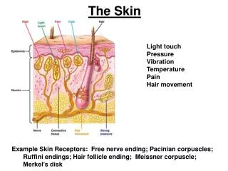
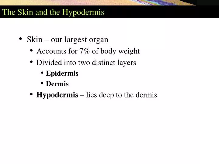
The Skin and the Hypodermis. Skin – our largest organ Accounts for 7% of body weight Divided into two distinct layers Epidermis Dermis Hypodermis – lies deep to the dermis. Skin Structure. Figure 5.1. The Skin and the Hypodermis. Functions Cushions and insulates deeper organs

E N D
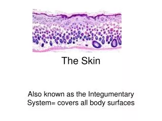
The Skin and the Hypodermis • Skin – our largest organ • Accounts for 7% of body weight • Divided into two distinct layers • Epidermis • Dermis • Hypodermis – lies deep to the dermis
Skin Structure Figure 5.1
The Skin and the Hypodermis • Functions • Cushions and insulates deeper organs • Protects body from bumps, scrapes, and cuts • Protects body from chemicals, heat, and cold (regulates body temperature) • Acts as a mini-excretory system • Screens out UV rays from the sun • Contains sensory receptors associated with nerve endings
Epidermal Cells and Layers of the Epidermis Figure 5.3
Epidermis • Contains four main cell types • Keratinocytes • Melanocytes • Merkel cells • Langerhans cells
Epidermis • Keratinocytes – most abundant cell type in epidermis • Arise from deepest layer of epidermis • Produce keratin – a tough fibrous protein • Produce antibodies and enzymes • Keratinocytes are dead at skin's surface • Merkel cells – associated with sensory nerve ending • Melanocytes – secrete the pigment melanin • Langerhans cells – innervate epidermal layers (communication)
Layers of the Epidermis • Stratum basale (stratum geminativum) • Stratum spinosum • Stratum granulosum • Stratum lucidum (only in thick skin) • Stratum corneum
Layers of the Epidermis • Stratum basale • Deepest layer of epidermis • Attached to underlying dermis • Cells actively divide • Stratum basale contains • Merkel cells – associated with sensory nerve ending • Melanocytes – secrete the pigment melanin
Layers of the Epidermis • Stratum spinosum (spiny layer) • "Spiny" appearance caused by artifacts of histological preparation • Contains thick bundles of intermediate filaments (tonofilaments) • Contains star-shaped Langerhans cells
Layers of the Epidermis • Stratum granulosum • Consists of keratinocytes and tonofilaments • Tonofilaments contain • Keratohyaline granules – help form keratin • Lamellated granules – contain a waterproofing glycolipid
Layers of the Epidermis • Stratum lucidum (clear layer) • Occurs only in thick skin • Composed of a few rows of flat, dead keratinocytes • Stratum corneum (horny layer) • Thick layer of dead keratinocytes and thickened plasma membranes • Protects skin against abrasion and penetration
Dermis • Second major layer of the skin • Strong, flexible connective tissue • Richly supplied with blood vessels and nerves • Has two layers • Papillary layer – includes dermal papillae • Reticular layer – deeper layer – 80% of thickness of dermis
Hypodermis • Deep to the skin – also called superficial fascia • Contains areolar and adipose connective tissues • Anchors skin to underlying structures • Helps insulate the body
Skin Color • Three pigments contribute to skin color • Melanin – most important pigment – made from tyrosine • Carotene – yellowish pigment from carrots and tomatoes • Hemoglobin – Caucasian skin contains little melanin • Allows crimson color of blood to show through
Sebaceous and Sweat Glands Figure 5.1
Sebaceous Glands • Occur over entire body, except palms and soles • Secrete sebum – an oily substance • Simple alveolar glands • Holocrine secretion – entire cell breaks up to form secretion • Most are associated with a hair follicle • Functions of sebum • Collects dirt; softens and lubricates hair and skin
Sweat Glands • Sweat glands (sudoriferous glands) widely distributed on body • Sweat – is a blood filtrate • 99% water with some salts • Contains traces of metabolic wastes
Sweat Glands • Two types of sweat gland • Eccrine gland • Most numerous – produce true sweat • Apocrine gland • Confined to axillary, anal, and genital areas • Produce a special kind of sweat
Burns • Classified by severity • First degree burn – only epidermis is damaged • Second degree burn – upper part of dermis is also damaged • Blisters appear • Skin heals with little scarring • Third degree burn – consume thickness of skin • Burned area appears white, red, or blackened
Estimating Burns Using the Rule of Nines Figure 5.11
Skin Cancer • Basal cell carcinoma – least malignant and most common • Squamous cell carcinoma – arises from keratinocytes of stratumspinosum • Melanoma – a cancer of melanocytes • The most dangerous type of skin cancer
Skin Cancer Figure 5.12