Download
1 / 134
1.36k likes | 1.51k Views
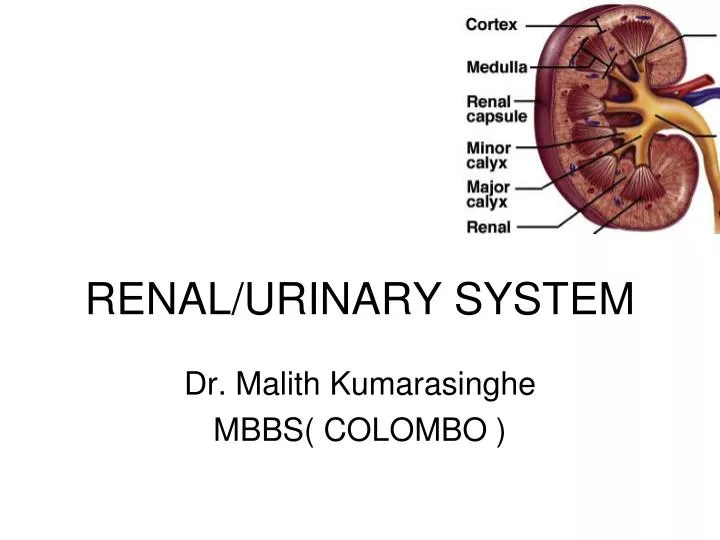
RENAL/URINARY SYSTEM. Dr. Malith Kumarasinghe MBBS( COLOMBO ). Functions of Urinary System. Two Kidneys carry out four functions Filter nitrogenous wastes, toxins, ions, etc. from blood to be excreted as urine. Regulate volume and chemical composition of blood (water, salts, acids, bases).

E N D
RENAL/URINARY SYSTEM Dr. Malith Kumarasinghe MBBS( COLOMBO )
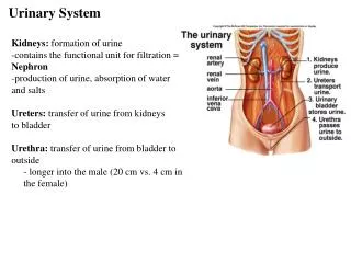
Functions of Urinary System • Two Kidneys carry out four functions • Filter nitrogenous wastes, toxins, ions, etc. from blood to be excreted as urine. • Regulate volume and chemical composition of blood (water, salts, acids, bases). • Produce regulatory enzymes. • Renin – regulates BP/ kidney function • Erthropoeitin – stimulates RBC production from marrow. • Metabolism of Vitamin D to active form
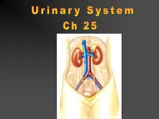
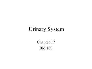
Two Ureters • Convey urine from Kidneys to Urinary Bladder • Urinary Bladder • Holds Urine until excretion • Urethra • Conveys urine from bladder to outside of body
Kidneys • Bean-shaped , reddish- brown organs • Lies retroperitoneal, on posterior abdominal wall • Lies from T12-L3 of vertebral column, next to m. psoas major • Superior parts are protected by ribs 11, 12 • Superior poles are closer to midline than inferior
Average size – 12cm x 6cm x 3 cm • Weights 150 grams
Right kidney lower than Left kidney WHY??????
External surface • Renal fascia (outer) • attaches kidney to posterior abdominal wall – flexible, allows kidney to move with respiration 2. Perirenal fat (middle layer) • protective cushion 3. Renal capsule (innermost) • layer of collagen fibres - barrier against trauma, infection etc
Kidney Internal Anatomy • Renal arteries and veins • Bring blood in and out of kidney • Renal cortex • Outer layer of Kidney • Renal medulla • Inner layer of Kidney
Kidney Internal Anatomy • Renal Pyramids • Renal Columns • Space between pyramids within the medula
Renal Papilla Narrow end of pyramid Calyx (ces) Collecting tubes Renal Pelvis Collecting vessel prior to ureter
Q3;Relations of Kidney? • Anterior • Postrior • Medial • Lateral
Ureters • Tubes that transport urine from renal pelvis to bladder • 20-30 cm long • Muscular walls - peristaltic waves force urine down to bladder
Retroperitoneal • Pressure in the bladder compresses ureter, helps prevent backflow of urine
Bladder • Hollow muscular organ • Retroperitoneal, posterior to pubic symphysis • Capacity ~ 300-400 ml (max = 1000 ml)
Empty: looks like a deflated balloon, rugae can be seen • Full spherical bladder rises above abdominal cavity • Trigone : triangular area bounded by openings of ureters and exit to urethra • Males: anterior to rectum, above prostate • Females: inferior to uterus, anterior to vagina
Relations • Anterior - pubic symphysis • Superior- Peritoneum( small intesine,sigmiod colon) • Lateral - levator ani, obturator internus • Posterior- male-rectum, seminal vesicles females-vagina, cervix
Support of bladder Martini p983 • superior surfaces - peritoneum • middle umbilical ligament • lateral umbilical ligaments • At base, tough ligamentous bands anchor bladder to pelvic and pubic bones
Cross section of ureter & bladder • Lined by stratified squamous epithelium to allow for stretch of wall • Transitional epithelium in the bladder
Smooth muscle (detrusor muscle) • inner and outer longitudinal • middle circular • Contraction compresses bladder and expels contents into urethra • Internal sphincter - involuntary control • External sphincter – voluntary control
Urethra Female ~ 4cm long • opens to exterior between clitoris and vaginal opening Male ~ 20 cm long • passes through prostate gland • pierces urogenital diaphragm • enters penis and extends throughout length • opens at urethral orifice
Nephron = Functional unit of the kidney, ~ 1 million nephrons per kidney Tubular components: 1. Glomerular (Bowman’s) capsule – double-walled cup • simple squamous epithelium 2. Proximal convoluted tubule- coiled 1st section • simple cuboidal epithelium with microvilli
3. Loop of Henle - hair-pin loop • thin descending limb, thick ascending limb 4. Distal convoluted tubule - last section • simple cuboidal epithelium • specialised region - Juxta glomerular apparatus Distal convoluted tubule opens into the collecting system collecting ducts papillary ducts minorcalyx…
Vascular component of nephron Made up of blood vessels: 1. Glomerulus - network of capillaries within Bowman’s capsule 2. Afferent arteriole - leading into glomerulus 3. Efferent arteriole - leading out of glomerulus 4. Peritubular capillaries - surrounding tubules 5. Vasa recta - specialised loops of blood vessels around long Loop of Henle (juxtamedullary nephrons)
5. Vasa recta - specialised loops of blood vessels around long Loop of Henle (juxtamedullary nephrons)
Cortical nephrons(85%) shorter, mostly in cortex of kidney, produce "standard" urine Juxtamedullary nephrons(15%), "juxta = next to" the medulla - responsive to ADH, can produce concentrated urine due to longer Loops of Henle Two Types of Nephrons
Q;6Draw a nephron • Vascular component • Tubular component
Consists of two structures: a glomerulus and a Bowman's capsule • FILTRATION APPARATUS OF KIDNEY BORMAN’S CAPSULE • Bowman’s capsule contains an inner visceral layer of epithelium around the glomerular capillaries and an outer parietal layer • simple squamous epithelium
The space between this two layers is continuous with the lumen of tubule and receives the glomerular filtrate GLOMERULUS • Specialized tuft of capillaries in the capsule (10-20 capillary loops) • Blood flowing through glomerulus capillaries undergoes a filtration process to produce the initial urine filtrate
FILTRATION MEMBRANE • 1- fenestrated capillaries; discontinuous endothelium consist of large pores • 2- continuous basal lamina • 3- podocytes of visceral layer of Bowman’s capsule; processes contact basal lamina and are separated by slits Glomerulus
Prevents RBC’s and large molecular weight proteins from leaving circulation, while most other blood constituents pass easily into the capsular space MESANGIAL CELLS • Phagocytic cells with a surrounding matrix that lend structural support to the glomerulus