Download
1 / 95
950 likes | 959 Views
Surface anatomy Plan fascia of the neck Dr . H.A.Jaafar Al- Nahrain University- college of Medicine Dept. Of Anatomy. Objectives: we should be able to know : The subcutaneous tissue nerves, veins of the neck,

E N D
Surface anatomy Plan fascia of the neck Dr. H.A.Jaafar Al-Nahrain University- college of Medicine Dept. Of Anatomy
Objectives: we should be able to know : The subcutaneous tissue nerves, veins of the neck, The main anatomical potential space in the neck which lead to Spread Infections to the mediastinum. Three major fascial compartments of the neck Where the viscera of the neck are located.
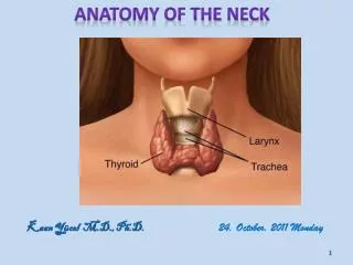
The Neck lies between lower margin of mandible above & suprasternal notch & upper border of clavicle below. It is strengthened by cervical part of vertebral column, is convex forward supports skull. Behind the vertebrae is ………………….a mass of extensor muscles in front is …………………………………..a smaller group of flexor muscles . In central region are :…………………parts of respiratory system, larynx & trachea, behind are parts of alimentary system, pharynx & esophagus. At sides of these structures are vertically running : carotid arteries, internal jugular veins, vagus nerve, deep cervical lymph nodes
Landmarks of the neck • Sternocleidomastoid • Suprasternal fossa • Greater supraclaviclar fossa
Landmarks of the neck • Hyoid bone • Thyroid cartilage • Cricoid cartilage
Skin of Neck lines of cleavage of skin are constant run …..horizontally …..around neck. an incision : along a cleavage line will heal as a narrow scar, crosses lines will heal as a wide or heaped-up scar.
The natural line of cleavage of the skin are constant and run almost horizontally around the neck
1-Superficial Fascia 2-deep Fascia
Superficial Fascia forms a thin layer encloses platysma muscle. embedded in it are : cutaneous nerves, superficial veins, superficial lymph nodes.
Structures in neck: are surrounded by a layer of subcutaneous tissue (superficial fascia) are compartmentalized by layers of deep cervical fascia. fascialplanes determine direction in which an infection in neck may spread. Cervical Subcutaneous Tissue &Platysma superficial cervical fascia is a layer of fatty connective tissue lies between dermis of skin & investing layer of deep cervical fascia It is usually thinner than in other regions, anteriorly. It contains : cutaneous nerves, blood & lymphatic vessels, superficial lymph nodes variable amounts of fat. Platysma……..Anterolaterally
external jugular vein (EJV) descending from angle of mandible to middle of clavicle are superficial to main cutaneous nerves of neck. covers anterolateral aspect of neck.
Platysma flat plate is a broad, thin sheet of muscle in subcutaneous tissue of neck is supplied by cervical branch of CN VII. Its fibers arise in deep fascia covering superior parts of deltoid & pectoralis major muscles sweep superomediallyover clavicle to inferior border of mandible. anterior borders of the two muscles decussate over chin blend with facial muscles. Inferiorly, fibers diverge, leaving a gap anterior to larynx & trachea
tenses skin, producing vertical skin ridges releasing pressure on superficial veins. use in shaving in a grimace. depress the mandible and draw corners of mouth inferiorly Acting its inferior attachment convey tension or stress.
Contents • Platysma • Superficial veins • Anterior jugular v. • External jugular v. • Cutaneous nerves • Lesser occipital n. • Greator auricular n. • Transverse nerve of neck • Supraclavicular n. • Cervical branch of facial n.
Regions of neck • Neck • Anterior region of neck • Sternocleidomastoid region • Lateral region of neck
Triangles of posterior (lateral) region of neck • Occipital triangle • supraclavicular triangle (greater supraclavicular fossa)
Deep Cervical Fascia: investing, pretracheal, & prevertebral.
support : viscera (thyroid gland), muscles, vessels, & deep lymph nodes. condenses around : ……… to form carotid sheath common carotid arteries, internal jugular veins (IJVs), & vagus nerves form natural cleavage planes tissues may be separated during surgery, limit the spread of abscesses (collections of pus) afford slipperiness allows structures in neck to move and pass over one another without difficulty, swallowing and turning the head and neck.
Deep Cervical Fascia: investing,
Investing Layer most superficial deep fascial layer, surrounds entire neck deep to skin and subcutaneous tissue. splits into superficial and deep layers to enclose (invest) : trapezius & sternocleidomastoid (SCM) muscles. Superiorly, attaches to : Superior nuchal line of occipital bone. Mastoid processes of temporal bones. Zygomatic arches. Inferior border of mandible. Hyoid bone. Spinous processes of cervical vertebrae. also splits to enclose : submandibular gland; posterior to mandible, it splits to form fibrous capsule of parotid gland.
Investing Layer stylomandibular ligament is a thickened modification Inferiorly, attaches to : manubrium, clavicles, & acromions spines of scapulae. continuous posteriorly with : periosteum covering C7 spinous process, nuchal ligament a triangular membrane forms a median fibrous septum between muscles of two sides of neck
Deep Cervical Fascia: pretracheal,
Pretracheal Layer of Deep Cervical Fascia is limited to the anterior part of neck It extends inferiorly from hyoid into thorax, it blends with the fibrous pericardium covering heart. includes a thin muscular part, encloses : infrahyoid muscles, & a visceral part, encloses thyroid gland, trachea, & esophagus pharynx is continuous posteriorly & superiorly with buccopharyngeal fascia of pharynx. blends laterally with carotid sheaths.
Pretracheal Layer of Deep Cervical Fascia In hyoid, a thickening of pretracheal fascia forms a pulley or trochlea through intermediate tendon of digastric muscle passes, suspending hyoid. tethers two-bellied omohyoid muscle, redirecting course of muscle between bellies.
forms a tubular sheath for vertebral column & muscles associated with it, such as : longus colli &longus capitis anteriorly, scalenes laterally, deep cervical muscles posteriorly is fixed to cranial base superiorly. Inferiorly, it blends with endothoracic fascia peripherally fuses with anterior longitudinal ligament centrally at approximately T3 vertebra extends laterally as axillary sheath surrounds axillary vessels & brachial plexus. sympathetic trunks cervical parts are embedded in it
is a tubular fascial investment extends from cranial base to root of neck. blends : anteriorly with investing and pretracheal layers posteriorly with prevertebral layer contains : common and internal carotid arteries, internal jugular vein, vagus nerve (CN X), deep cervical lymph nodes, carotid sinus nerve, sympathetic nerve fibers (carotid periarterial plexuses). carotid sheath and pretracheal fascia communicate freely with: mediastinum of thorax inferiorly & cranial cavity superiorly. represent potential pathways for spread of infection and extravasated blood.
Investing layer of deep cervical fascia Sternocleidomastoid Pretrachealfascia (visceral part) Carotid sheath T Pretracheal fascia (muscular part) E Alar fascia Buccopharyngeal fascia Prevertebral fascia Trapezius Deep Cervical Fascia is largest and most important interfascial space in neck It is a potential space consists of loose connective tissue between visceral part of prevertebral layer of deep cervical fascia & buccopharyngeal fascia surrounding pharynx superficially. Inferiorly, buccopharyngeal fascia is continuous with pretracheal layer alar fascia : forms a further subdivision of retropharyngeal space. is attached along midline of buccopharyngeal fascia from cranium to level of the C7 vertebra. it extends laterally and terminates in carotid sheath. permits movement of pharynx, esophagus, larynx, and trachea relative to vertebral column during swallowing. is closed : superiorly by cranial base and on each side by carotid sheath. It opens inferiorly into superior mediastinum
Posterior cervical triangle Investing layer of deep cervical fascia Sternocleidomastoid Trapezius Pretrachealfascia (visceral part) Carotid sheath T Pretracheal fascia (muscular part) E Alar fascia Buccopharyngeal fascia Sternocleidomastoid Prevertebral fascia Trapezius Deep Cervical Fascia Anterior triangles Suprahyoidmuscles Infrrahyoidmuscles Prevertebral muscles Scalene muscles
Sup. thyroid Ext. jugular Int. jugular Middle thyroid Inf. thyroid Ant. jugular Trace the pathways for venous drainage from the neck into the brachial veins.
Lesser occipital n. External jugular vein Greet auricular n. Transverse nerve of neck Supraclavicular n. Anterior jugular vein Cutaneous nerves and superficial veins
Submendibular gland Digastric Accessory n. Hypoglossal n. Superior thyroid a. Ansa cervicalis Cervical plexus Sternothyroid Phrenic n. Sternohyoid Omohyoid Vagus n.