Download
1 / 13
140 likes | 306 Views
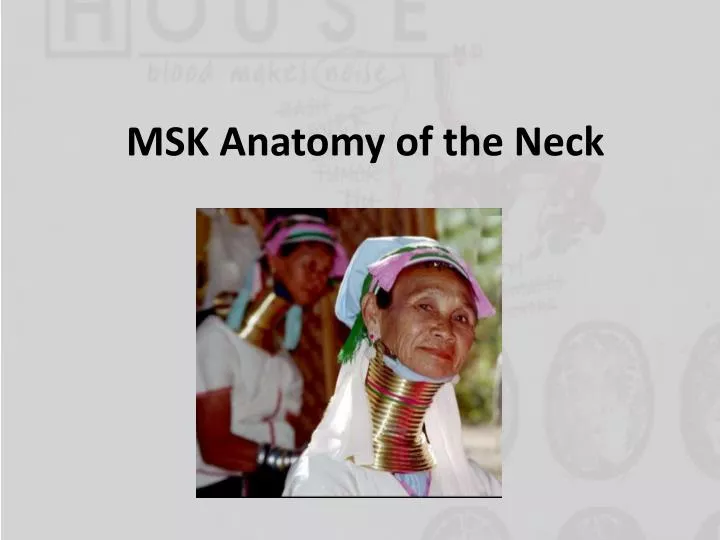
MSK Anatomy of the Neck. Osteology of the Vertebral Column. Vertebrae. Vertebral body (general plan):. Intervertebral disc:. Cervical Vertebrae. Atlas (C1):. Typical:. Thoracic Vertebrae. Lumbar Vertebrae. Vertebral Ligaments. Ligamentum flavum : joins up vertebra via lamina

E N D
Vertebrae Vertebral body (general plan): Intervertebral disc:
Cervical Vertebrae Atlas (C1): Typical:
Vertebral Ligaments • Ligamentumflavum: joins up vertebra via lamina • Ligamentumnuchae: same as supraspinous ligament (but in neck) • If you wanted to get CSF, you would penetrate these layers (in order) • Skin Subcutaneous tissue Supraspinous ligament Interspinous ligament Ligamentumflavum Dura mater Subarachnoid mater Subarachnoid space
Spinal Cord • Layers in the spinal cord: • Dura mater (tough) • Arachnoid mater (web-like) • Pia mater (delicate)
Posterior Triangle • A fascial carpet covers five muscles: • Splenius capitis • Levator scapulae • Scalenus posterior • Scalenusmedius • Scalenus anterior


![[PDF] Illustrated Anatomy of the Head and Neck Kindle](https://cdn7.slideserve.com/12506909/slide1-dt.jpg)
![[PDF] DOWNLOAD Illustrated Anatomy of the Head and Neck](https://cdn7.slideserve.com/12519396/slide1-dt.jpg)