Download
1 / 24
590 likes | 1.58k Views
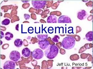
Leukemia. The leukemias are the most common malignant neoplasms in childhood: 41 % of all malignancies that occur in children less than 15 yrs. 77% - Acute lymphoblastic leukemia (ALL) 77 %. 11% - Acute myelogenous leukemia (AML ) 2-3% - Chronic myelogenous leukemia (CML )

E N D
Leukemia • The leukemias are the most common malignant neoplasms in childhood: • 41% of all malignancies that occur in children less than 15 yrs. • 77% - Acute lymphoblastic leukemia (ALL) 77%. • 11% - Acute myelogenous leukemia (AML) • 2-3% - Chronic myelogenous leukemia (CML) • 1-2% - Juvenile chronic myelogenous leukemia (JCML)
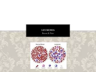
Leukemia • A group of malignant diseases in which genetic abnormalities in a hematopoietic cell give rise to a clonal proliferation of cells. • Increased rate of proliferation, a decreased rate of spontaneous apoptosis, or both. • Disruption of normal marrow function and, ultimately, marrow failure.
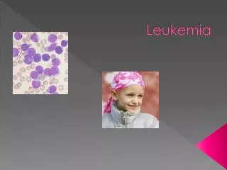
Acute Epidemiology: • Peak incidence between 2–6 yr of age. • Male predominance. • More common in children with certain chromosomal abnormalities such as Down syndrome, Bloom syndrome, ataxia-telangiectasia, and Fanconi syndrome.
Etiology: • The etiology of ALL is unknown, although several genetic and environmental factors are associated with childhood leukemia. • Exposure to medical diagnostic radiation both in utero and in childhood. • Association between B-cell ALL and Epstein-Barr viral infections in certain developing countries.
Genetic Conditions Predisposing to Childhood Leukemia • Down syndrome • Fanconi syndrome • Bloom syndrome • Diamond-Blackfan anemia • Schwachman syndrome • Klinefelter syndrome • Turner syndrome • Neurofibromatosis • Ataxia-telangiectasia • Severe combined immune deficiency • Paroxysmal nocturnal hemoglobinuria • Li-Fraumeni syndrome
Environmental Factors Predisposing to Childhood Leukemia • Ionizing radiation • Drugs • Alkylating agents • Nitrosourea • Epipodophyllotoxin • Benzene exposure • Advanced maternal age
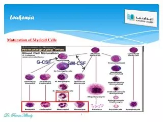
Pathogenesis • The classification of ALL depends on characterizing the malignant cells in the bone marrow to determine the morphology, phenotypic characteristics as measured by cell membrane markers, and cytogenetic and molecular genetic features. • Morphology alone is usually adequate to establish a diagnosis, but the other studies are essential for disease classification, which may have a major influence on both the prognosis and the choice of appropriate therapy.
Pathogenesis • Chromosomal abnormalities are found in most patients with ALL. • These abnormalities provide important prognostic information. • PCR and fluorescence in situ hybridization techniques offer the ability to pinpoint molecular genetic abnormalities and to detect small numbers of malignant cells during follow-up.
Acute Lymphoblastic Leukemia French-American-British classification
Clinical Manifestations • The initial presentation of ALL is usually nonspecific and relatively brief: • Anorexia • Fatigue • Irritability • Intermittent low-grade fever • Bone or joint pain, particularly in the lower extremities, may be present. • Patients often have a history of an upper respiratory tract infection in the proceeding 1–2 mo.
Clinical Manifestations • symptoms may be of several months duration and may be predominantly localized to the bones or joints • As the disease progresses, signs and symptoms of bone marrow failure become more obvious: • pallor, fatigue, bruising, epistaxis, fever that may be caused by infection Physical examination reflecting bone marrow failure: • Pallor • purpuric and petechial skin lesions • mucous membrane hemorrhage
Clinical Manifestations • The proliferative nature of the disease may be manifested as • Lymphadenopathy • Splenomegaly • Hepatomegaly • Signs of increased intracranial pressure that indicate leukemic involvement of the CNS: • Papilledema • Retinal hemorrhages • Cranial nerve palsies
Diagnosis • Strongly suggested by peripheral blood findings indicative of bone marrow failure. • Anemia and thrombocytopenia are seen in most patients. • Leukemic cells are often not observed in the peripheral blood in routine laboratory examinations. • Most patients with ALL present with total leukocyte counts of less than 10,000/μL
Diagnosis • When the results of an analysis of peripheral blood suggest the possibility of leukemia, a bone marrow examination should be done promptly to establish the diagnosis. • Bone marrow aspiration alone is usually sufficient, but sometimes a bone marrow biopsy is needed to provide adequate tissue for study or to exclude other possible causes of bone marrow failure.
Diagnosis • ALL is diagnosed by a bone marrow evaluation that demonstrates more than 25% of the bone marrow cells as a homogeneous population of lymphoblasts. • Staging of ALL is partly based on a CSF examination. • If lymphoblasts are found and the CSF leukocyte count is elevated, overt CNS leukemia is present.
Treatment • Three of the most important predictive factors are: • Age of the patient • Initial leukocyte count • The speed of response to treatment • Patients between 1–10 yr of age and with a leukocyte count of less than 50,000/μL is widely used to define average risk. • Patients considered to be at higher risk are children who are older than 10 yr of age or who have an initial leukocyte count of more than 50,000/μL.
Treatment • The outcome for patients at higher risk can be improved by administration of more intensive therapy despite the greater toxicity of such therapy. • Infants with ALL, along with patients who present with specific chromosomal abnormalities such as t(9;22) or t(4;11), have an even higher risk of relapse despite intensive therapy. • The prognosis for patients with a slower response to initial therapy may be improved by therapy that is more intensive than the therapy considered necessary for patients who respond more rapidly.
Treatment • The initial therapy is designed to eradicate the leukemic cells from the bone marrow and is known as remission induction. • During this phase, therapy is usually given for 4 wk and consists of: • Vincristineweekly. • Corticosteroid such as dexamethasone or prednisone. • Either repeated doses of native L-asparaginaseor a single dose of a long-acting asparaginase preparation. • Intrathecalcytarabine or methotrexate, or both, may also be given.
Treatment • With this approach, 98% of patients are in remission, defined as <5% blasts in the marrow and return of neutrophil and platelet counts to near normal levels after 4-5weeks of treatment.
Treatment • The second phase of treatment focuses on CNS therapy in an effort to prevent later CNS relapses. • Intrathecalchemotherapy is given repeatedly by lumbar puncture in conjunction with intensive systemic chemotherapy. • The likelihood of later CNS relapse is thereby reduced to less than 5%. • A small proportion of patients with features that predict a high risk of CNS relapse receive irradiation to the brain and spinal cord: • This includes those patients who have lymphoblasts in the CSF and an elevated CSF leukocyte count at the time of diagnosis.
Treatment • After remission induction, many regimens provide 14–28 wk of multiagent therapy • Finally maintenance phase of therapy, lasts for 2–3 years depending on the protocol used: • Daily mercaptopurine and weekly methotrexate, usually with intermittent doses of vincristine and a corticosteroid. • Poor prognostic features: • Those with the t(9;22) translocation may undergo bone marrow transplantation during the first remission.
Treatment • The major impediment to a successful outcome is relapse of the disease. • Relapse occurs in the bone marrow in 15–20% of patients with ALL and carries the most serious implications, especially if it occurs during or shortly after completion of therapy. • Intensive chemotherapy with agents not previously used in the patient followed by allogeneic stem cell transplantation can result in long-term survival for a few patients with bone marrow relapse.
Acute Lymphoblastic Leukemia • SUPPORTIVE CARE: • -Close attention to the medical supportive care needs of the patients is essential in successfully administering aggressive chemotherapeutic programs. • -Patients with a large tumor burden are prone to tumor lysis syndrome • -Chemotherapy may produce severe myelosuppression, • may require erythrocyte and platelet transfusion • always requires a high index of suspicion and aggressive empirical antimicrobial therapy for sepsis in febrile children with neutropenia. • -prophylactic treatment of Pneumocystiscarinii pneumonia during chemotherapy and for several months after
Acute Lymphoblastic Leukemia • Prognosis: • -Most children with ALL can now be expected to have long-term survival, with the rate greater than 80% after 5 yr. • -The most important prognostic factor is the choice of appropriate risk-directed therapy, with the type of treatment chosen according to the type of ALL, the stage of disease, the age of the patient, and the rate of response to initial therapy. • -Characteristics generally believed to adversely affect outcome include: • an age younger than 1 yr or older than 10 yr at diagnosis. • a leukocyte count > 100,000/μL at diagnosis. • a slow response to initial therapy.