Download
1 / 61
610 likes | 804 Views
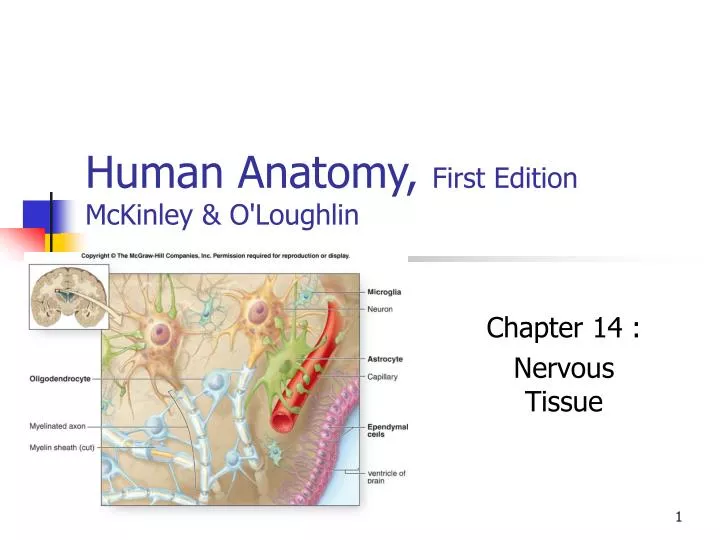
Human Anatomy, First Edition McKinley & O'Loughlin. Chapter 14 : Nervous Tissue. The Nervous System. The body’s primary communication and control system. Can be divided according to: Structural categories Functional categories. Nervous System: Structural Organization.

E N D
Human Anatomy, First EditionMcKinley & O'Loughlin Chapter 14 : Nervous Tissue
The Nervous System • The body’s primary communication and control system. • Can be divided according to: • Structural categories • Functional categories.
Nervous System: Structural Organization Structural subdivisions of the nervous system: • Central nervous system (CNS) • brain and spinal cord • Peripheral nervous system (PNS) • cranial nerves (nerves that extend from the brain) • spinal nerves (nerves that extend from the spinal cord) • ganglia (clusters of neuron cell bodies (somas) located outside the CNS)
Nervous System: Functional Organization Functional divisions of the nervous system: • Sensory afferent division: • receives sensory information (input) from receptors • transmits this information to the CNS. • Motor efferent division: • transmits motor impulses (output) from the CNS • to muscles or glands (effector organs).
Sensory Division: two components • Somatic sensory components: • General somatic senses: • touch • pain • pressure • vibration, • temperature • proprioception. • Special senses: • Taste • Vision • Hearing • Balance • smell
Sensory Division: two components • Visceral sensory components • transmit nerve impulses from blood vessels and viscera to the CNS • visceral senses primarily include: • temperature • stretch (of the organ wall).
Motor Division: two components • The somatic motor component (somatic nervous system; SNS): • conducts nerve impulses from the CNS to skeletal muscles • also known as the voluntary nervous system • The autonomic motor component (autonomic nervous system; ANS): internal organs, regulates smooth muscle, cardiac muscle, and glands. • Innervates • Internal organs • Regulates smooth muscle • Regulates cardiac muscle • Regulates glands • also known as the visceral motor system or involuntary nervous system
Nerve Cells • Nervous Tissue • Two distinct cell types • Neurons • excitable cells • initiate and transmit nerve impulses • Glial cells • nonexcitable cells • support and protect the neurons
Characteristics of Neurons • Neurons have a high metabolic rate. • Neurons have extreme longevity. • Neurons typically are non-mitotic.
Neuron Structure • Neurons come in all shapes and sizes • All neurons share certain basic structural features. • typical neuron: • Cell body (soma, perikaryon) • Dendrites • Axon • Collaterals: branches • axon terminals or telodendria • Synaptic knobs
Neuron Structure – Cell Body • The cell body (perikaryon, soma) • the neuron’s control center • responsible for: • receiving • integrating • sending nerve impulses. • Consists of: • Plasma membrane • Cytoplasm • Nucleus with prominent nucleolus • Chromatophobic substance (Nissil bodies): RER • Free ribosomes
Neuron Structure – Dendrites • Shorter, smaller processes • Branch off the cell body. • Some neurons have only one dendrite, while others have many. • Dendrites conduct nerve impulses toward the cell body • they receive input • transfer input to the cell body for processing. • The more dendrites a neuron has, the more nerve impulses that neuron can receive from other cells.
Neuron Structure – Axon • larger, typically longer nerve cell process • Extend from the cell body • Axon hillock • also calleda nerve fiber • Most neurons have only one axon. • Anaxonic
Neuron Structure – Axon • Structures • Collaterals • Telodendria (axon terminals) • Synaptic knobs (terminal boutons) • The axon transmits a nerve impulse away from the cell body toward another cell.
Neuron Structure • Cytoskeleton • Neurotubules • microtubules • Neurofilaments • Intermediate fibers • Neurofibrils • Bundles of neurofibrils • In both dendrites and axons • Provide strength
Classifications of Neurons • Neurons vary widely in morphology and location. • classified based on • structure • function. • Structural classification: number of processes extending from the cell body. • unipolar neuron has a single process • bipolar neurons have two processes • multipolar neurons have three or more processes
Functional Classification • Sensory afferent neurons: receptor to CNS • Motor efferent neurons: CNS to effector • Interneurons (association neurons): facilitate communication between sensory and motor neurons.
Interneurons • Interneurons, or association neurons • lie entirely within the CNS • multipolar. • They receive nerve impulses from many other neurons • They carry out the integrative function of the nervous system. • Interneurons facilitate communication between sensory and motor neurons.
Glial Cells • Also called neuroglia • Occur within both the CNS and the PNS. • are smaller than neurons • are capable of mitosis. • do not transmit nerve impulses. • Glial cells • physically protect neurons • help nourish neurons • provide a supporting framework for all the nervous tissue. • Glial cells far outnumber neurons. • Glial cells account for about half the volume of the nervous system.
Glial Cells of the CNS: astrocytes • Exhibit a starlike shape due to projections from their surface. • The most abundant glial cells in the CNS • constitute over 90% of the tissue in some areas of the brain. • Help form the blood-brain barrier (BBB): • strictly controls substances entering the nervous tissue in the brain from the bloodstream. • Regulate tissue fluid composition. • Provide structural support • Replace damaged neurons • Assist neuronal development
Glial Cells of the CNS: ependymal cells • Cuboid ET • Cilia on apical surface • Circulates CSF. • Line internal cavities • Processes make contact with other glial cells • Help form the choroid plexus • CSF: cerebral spinal fluid
Glial Cells of the CNS: microglia • Smallest % of CNS glial cells. • Phagocytic • Move through the tissue in response to infection • Remove debris. • Like macrophages
Glial Cells of the CNS: oligodendrocytes • Large, with big body and processes. • Processes form myelin sheaths • Speeds up transmission
Glial Cells of the PNS • Satellite cells: • Flattened cells • Cover somas in ganglia • Separate soma from surrounding tissue fluid • Regulate exchange. • Neurolemmocytes (Schwann cells) • Myelination in the PNS
Myelination • Process by which part of an axon is wrapped with a myelin sheath • Forms a protective fatty coating • Has a glossy-white appearance. • The myelin sheath: • supports the axon • protects the axon • insulates an axon
Myelination • No change in voltage can occur across the membrane in the insulated portion of an axon. • Voltage change occurs at the nodes • Neurolemmocytes: form myelin sheaths in PNS • Oligodendrocytes: form myelin sheaths in the CNS
Mylenated vs. Unmylenated Axons • myelinated axon • nerve impulse “jumps” from neurofibril node to neurofibril node • known as saltatory conduction • requires less energy (ATP) than does an unmyelinated axon • unmyelinated axon • nerve impulse must travel the entire length of the axon • known as continuous conduction • nerve impulse takes longer to reach the end of the axon • Using continuous conduction, unmyelinated axons conduct nerve impulses from pain stimuli • A myelinated axon produces a faster nerve impulse.
Regeneration of PNS Axons • PNS axons are vulnerable to cuts and trauma. • A damaged axon can regenerate • if some neurilemma remains. • PNS axon regeneration depends upon three factors. • amount of damage • neurolemmocyte secretion of nerve growth factors • stimulates outgrowth of severed axons • distance between the site of the damaged axon and the effector organ
Regeneration of PNS Axons • Wallerian degeneration. • Axon damaged • Proximal end seals, and swells. • Distal end degenerates, macrophages clean up • Distal neurolemmocytes survive • Neurolemmocytes form regeneration tube (with endoneurinum) • Axon regenerates, remyelinates • Axon reestablishes contact with effector
Structure of a Nerve • A nerve is a cable-like bundle of parallel axons. • three connective tissue wrappings. • Endoneurium • delicate layer of loose connective tissue • Perineurium • a cellular and fibrous connective tissue layer • wraps groups of axons into fascicles • Epineurium - a superficial connective tissue covering • This thick layer of dense irregular fibrous connective tissue • encloses entire nerve • provides support and protection
Nerves • Nerves are organs of the PNS. • Sensory (afferent) nerves convey sensory information to the CNS. • Motor (efferent) nerves convey motor impulses from the CNS to the muscles and glands. • Mixed nerves: both sensory and motor • Axons terminate as they contact other neurons, muscle cells, or gland cells. • An axon transmits a nerve impulse at a specialized junction with another neuron called synapse.
Synapses • Presynaptic neurons • transmit nerve impulses toward a synapse. • Postsynaptic neurons • conduct nerve impulses away from the synapse. • Axons may establish synaptic contacts with any portion of the surface of another neuron • except those regions that are myelinated.