Download

1 / 13
130 likes | 307 Views
Tetrahymena as a model system to study phagocytosis. Module 2 – Week 1. Before we start this week…. We have assigned new 4-person lab groups for everyone You will be doing much of the work in pairs today Half of class will start exercise #1: pipetting

E N D
Tetrahymena as a model system to study phagocytosis Module 2 – Week 1
Before we start this week… • We have assigned new 4-person lab groups for everyone • You will be doing much of the work in pairs today • Half of class will start exercise #1: pipetting • Other half will start exercise #2: microscopy
Goals 1) Become competent in the use of micropipettors to deliver very small volumes of liquids. 2) Develop the ability to use the Olympus CH-2 brightfield microscope for studying single-celled organisms. Next week… 3) Explore how polystyrene microbeads can be used to identify subcellular structures and organelles. 4) Develop an experimental strategy to test a hypothesis related to the phagocytotic mechanism of Tetrahymena.
Gilson Pipetman • Identity of pipet on top button • P20 and P200 use yellow tips • P1000 uses blue tips • Two stop positions on piston • 1st: to fill • 2nd: to expel remaining liquid during dispensing • The numbers on the dial depends on the pipet in use
Pipetting exercise • P-1000 pipettorset to 500 μl (water weight = 500 mg = 0.500 g) • P200 set to 200 μl (water weight = 200 mg) • P20 set to 20 μl (water weight = 20 mg) • Empty 1.7 ml centrifuge tube on a 3-place pan balance. Tare (zero) the balance so the weight of the tube is zeroed out. • Pipette water into the tube. Weigh the tube and record the weight. • Repeat five times. • Repeat this exercise with all three pipets. • Using Excel, calculate the average and standard deviation of your three different pipettors.
Introduction to the microscope • Place sample slide with coverslip facing up on specimen holder. • Turn the light intensity dial to the zero position. • Turn the main power switch on and increase the light level. • Adjust specimen stage so that edge of glass is in beam. • Make sure the condenser is all the way down. • Rotate the 10x objective into position for viewing. • Turn the coarse focus knob to a position so that the specimen is basically in focus. • Adjust the distance between two eyepieces (interpupillary distance) so that you see through both eyes. • Use the fine-focus knob to achieve optimum focus through the right ocular. • Use the diopter adjustment ring to focus the image through the left ocular, so that both eyes see the image properly focused. • Adjust the aperture iris (using its lever) for optimum contrast. The iris aperture must be adjusted each time you switch to a different objective. • After focusing with the 10x objective, you can increase total magnification with the 40x objective. You will need to adjust the fine-focus each time you change. • Don’t let any objective lens contact the slide. • Adjust the light intensity when you change objectives, and then re-adjust the iris aperture.
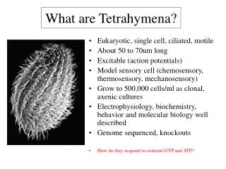
Viewing Tetrahymena in the microscope • Tetrahymena cell culture prepared for you • Three 1.7 ml microtubes: A, B, and C. • A = 50 μl of Tetrahymena + 50 μlof 3 micron polystyrene beads. • B = 50 μl of Tetrahymena + 50 μl of 0.2% glutaraldehyde. • PERFORM IN FUME HOOD; CAUTION: glutaraldehyde is highly toxic. • Mix both tubes gently. • C = 20 μl of cells from Tube “A” (~10 min bead exposure) + 20 μl of glutaraldehyde, then mix. Record the duration of ink exposure. • PERFORM IN FUME HOOD • Observe three Tetrahymena samples in the microscope
Glutaraldehyde • Hazard Statements: • H302: Harmful if swallowed • H315: Causes skin irritation • H317: May cause an allergic skin reaction • H318: Causes serious eye damage • H330: Fatal if inhaled • H334: May cause allergy or asthma symptoms or breathing difficulties if inhaled • H400: Very toxic to aquatic life • If you would like to review the complete MSDS, please consult with your TA
Proper Personal Protective Equipment (PPE) • All Glutaraldehyde must be handled in the fume hood • Every student must wear a lab coat, gloves, and safety glasses when working with 2.5% glutaraldehyde or samples treated with glutaraldehyde
Disposal of Tips, Slides, and Tubes • Tips that have not come in contact with glutaraldehyde can be placed in the coffee can labeled “Tips Only” located on your lab bench. • Slides that have not been contaminated with glutaraldehyde can be put in the red “sharps” container on the side bench. • All tubes containing liquid with trace glutaraldehyde must be emptied into the container in the fume hood labeled “Liquid Waste.” • Tips, tubes, and slides contaminated with glutaraldehyde must go in the container in the fume hood labeled “Solid Waste.” • Gloves and kimwipes must go in the Biohazard Box located by the hood • Wipe down your bench before leaving and put paper towel in Biohazard Box • Don’t touch computers with gloved hands
Write up your work today • Pipetting exercise • Calculate mean and S.D. for each pipet verification • Determine whether pipetting technique was precise or imprecise • Initial observations of Tetrahymena • Drawing of typical cell • Describe swimming and eating behaviors • Effects of glutaraldehyde • For fixed cells exposed to beads, count number of beads per cell for ten cells. Mean and S.D. • Turn in your observations, results and conclusions for Exercise #1 and #2 next lab session.
Next week’s experiment? • From your initial observations, consider exploring the following: • What do Tetrahymena consider food? • What is the cellular mechanism by which food is ingested? • ??? • Fill out the “My Proposed Experiment” form and turn in with summary of today’s work on flip side