Download
1 / 19
200 likes | 363 Views
about mechanism of phagocytosis and new findings on phagocytosis

E N D
PHAGOCYTOSIS PRESENTATION BY: BHANU SHRESTHA MSC FIRST SEMESTER MICROBIOLOGY
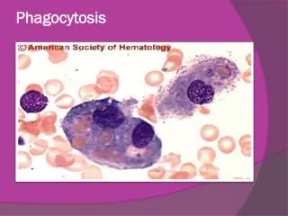
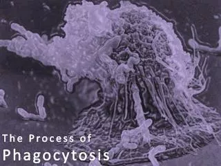
Background In cell biology, phagocytosis (from Ancient Greekφαγεῖν (phagein) , meaning "to devour", κύτος, (kytos) , meaning "cell", and -osis, meaning "process") is the process by which a cell—often a phagocyte or a protist—engulfs a solid particle to form an internal vesicle known as a phagosome. Phagocytosis was first noted by Canadian physician William Osler, and later studied by Élie Metchnikoff. Phagocytosis is a type of endocytosis. Endocytosis is a process through which a cell absorbs a particle, molecule, bacterium, or other type of matter by engulfing it. Phagocytosis refers to the engulfing of larger, solid particles. Often the engulfed particle is another cell, like when a white blood cell, which is a part of the immune system, engulfs a bacterium to destroy it. Phagocytosis is second line of defence mechanism which acts as cellular and humoral barrier. Professional phagocytes, such as macrophages, neu-trophils and dendritic cells, are uniquely qualified to engulf large (≥ 0.5 μm) particles, including microorgan-isms
Steps of phagocytosis • Chemotaxis • Attachment • Ingestion • Intracellular killing • Egestion
1.Chemotaxis • Chemotaxis is the process whereby recognition of an external gradient induces polarized actin polymerization resulting in persistent and directional cell body translocation. • Binding of chemoattractantligand activates a signal transduction cascade that promotes the formation of a pseudopod at the “front” of the cell and subsequent retraction of the midbodyanteriorly, resulting in “forward” cell motility. • Resting phagocytes are activated by inflammatory mediators and produce surface receptors that increase their ability to adhere to the inner surface of capillary walls enabling them to squeeze out of the capillary and enter the tissue, a process called diapedesis. Activation also enables phagocytes to produce endocytic pattern-recognition receptors that recognize and bind to microbial PAMPs in order to attach the microbe to the phagocyte, as well as to exhibit increased metabolic and microbicidal activity. Phagocytes then use chemotaxis to move towards an increasing concentration of some attractant such as bacterial factors or defense molecules. • New finding: Many results demonstrated the abilities of PS-G(the polysaccharide from Ganodermalucidum ) to enhance neutrophil function in phagocytosis and chemotaxis, and further provide evidence to strengthen the beneficial remedy of G. lucidum In human to enhance defense system.
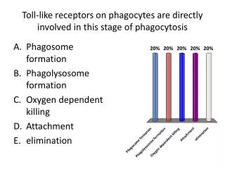
2.Attachment • The white blood cell then has to attach its membrane to the membrane of the bacterium. It does this by using molecules called surface receptors. These are molecules embedded in the white blood cell's membrane that are designed to detect and attach to molecules in the membrane of the bacterium. The two cell membranes link up and stick together. • Attachment of phagocytes to the microbes or cells can be through unenhanced attachment or enhanced attachment. Unenhanced attachment is the recognition of pathogen-associated molecular patterns or PAMPs by endocytic pattern-recognition receptors on the surface of the phagocytes. Enhanced attachment, or opsonization, is the attachment of microbes to phagocytes by way of an antibody molecule called IgG, the complement proteins C3b and C4b, and acute phase proteins such as mannose-binding lectin (MBL) and C-reactive protein (CRP).
3.Igestion • Once attached to each other, the membrane of the white blood cell swells outward around the bacterium and engulfs it. The membrane enclosing the bacterium pinches off and the result is a little pouch, called a phagosome, that contains the offending bacterium inside of the white blood cell. • Following attachment, polymerization and then depolymerization of actin filaments send pseudopods out to engulf the microbe and place it in an endocytic vesicle called a phagosome.During this process, an electron pump brings protons (H+) into the phagosome to lowers the pH within the phagosome to a pH that is correct for the acid hydrolases to effectively break down cellular proteins.
4.Intracelluar killing • Phagocytes contain membranous sacs called lysosomes that contain various digestive enzymes, microbicidal chemicals, and toxic oxygen radicals. The lysosomes fuse with the phagosomes containing the ingested microbes and the microbes are destroyed. • Intracellular killing mechanism by phagocytosis is from 3 methods which depend upon the nature of pathogen and they are as follows: • (A) O₂-dependent mechanism • (B) O₂-independent mechanism • (C) Nitrogen -dependent mechanism
(A) O₂-dependent mechanism • This mechanism is known as oxygen burst. • Activation of macrophage activates membrane bound oxidase enzyme. • These enzymes catalyzes reduction of oxygen to superoxide anion • These superoxides anions are toxic to ingested microorganism • The superoxide anions also generates other powerful oxidising agents such as hydroxyl radical and H₂O₂. • As the lysosome fuses with the phagosome, myeloperoxidase enzyme activates which produces hypochlorite from H₂O₂. • Hypochlorite is toxic to ingested microorganism. • O₂ + e⁻ O₂⁻ (mediated by oxidase) • 2 O₂⁻ + 2H⁺ H₂O₂ + O₂ • H₂O₂ + Fe⁺⁺ OH• + OH⁻ + Fe⁺⁺⁺ • H₂O₂ + Cl⁻ HOCl(ClO⁻) (mediated by Myeloperoxidase)
(B) O₂-independent mechanism • Activated macrophage activates different types of lytic enzymes inside the phagolysosome. • Lytic enzymes are: 1.Lysosome 2.Hydrolytic enzymes 3.Defensins (1)Lysossome: • Lysosomes are basically sacs containing digestive enzymes. • Lysosomes are used by the cell to digest or breakdown multifaceted organic molecules.Theses include proteins, fats, polysaccharides, and nucleic acids. (2)Hydrolytic enzymes: • They are elastase,collagenase,protease,acidhydrolase. • Protease is further divided into shearing protease(e.g. elastase) and metallo protease(e.g.collagenase,gellatinase etc.)
(3)Defensins: • It is antimicrobial and cytotoxic peptides. • It is cysteineriched cationic peptide that contains 29-35 amino acid residues. Each peptide contain invariants cysteins that forms a circular molecule. • These circular molecule are stabilised by intramolecular disulphide bond. • These circular defensins form ion permeable channels in bacterial cell membrane. • Defensins can kill ingested S. aureus, Streptococcus pneumoniae, E. coli, Pseudomonas aerogenesa, Haemophilus influenza.
(C) Nitrogen-dependent mechanism: • When macrophages are activated with bacterial cell wall componenets such as lipopoplysaccharides(gram negative) or in the case of Mycobacteria muramyl dipeptide(MDP) • Together with signalling molecule interferon γ results in activation of high level of nitric oxide synthetase enzymes inside the phagolysosome. • This enzymes catalyzes the conversion of L-arginine to nitric oxide by nitric oxide synthetase. • Nitric oxide has potent antimicrobial activity. • It can also combine with the superoxide anion and produce even more potent antimicrobial substances(hydroxyl radical peroxynitrites) (nitric oxide synthetase) L-arginine+ O₂ + NADPH NO + L-citrulline + NADP (nitric oxide)
5.Egestion • Macrophages and monocytes live much longer than neutrophils and must dispose of their bacterial components. Once microorganisms are destroyed, the unwanted organic material is expelled from the cell in a process called egestion. Egestion is the opposite of ingestion and the molecular mechanism is basically the reverse of phagocytosis with the microbial leftovers being dumped into the blood and lymph. Some of this microbial debris is not egested, but binds to special protein complexes (called Major Histocompatibility Complex molecules) on the membranes of macrophages for presentation to the immune system.
Factors affecting SP-A-mediated phagocytosis in human monocytic cell lines. • SP-A(Surfactant protein-A) derived from human lavage(bronchoalveolar washing) appeared to enhance phagocytosis by two different mechanisms; 1. by SP-A binding of the target to enhance its recognition and subsequent phagocytosis 2. by a direct SP-A stimulatory effect on the phagocyte itself. • Both SP-A mechanisms occurred with different targets in the same experimental system and the SP-A effects were qualitatively (but not quantitatively) comparable in several human cell lines. It also found that the SP-A effects were abrogated(repealed) when SP-A was combined with surfactant lipids, but the lipids did not affect the basal level of phagocytosis or phagocytosis by mechanisms not involving SP-A. Moreover, the stimulatory effect of SP-A was pH-dependent and appeared to be independent of several other phagocytic mechanisms, including those mediated by Fc receptors and mannose receptor.