Download
1 / 23
460 likes | 4.55k Views
CRP vs. ESR Assessing and Measuring the Inflammatory Response.

E N D
CRP vs. ESRAssessing and Measuring the Inflammatory Response
ESR and CRP are some of the most commonly requested tests by New Zealand general practitioners. ESR is being performed about twice as often as CRP, but it is now recognised that in most situations CRP provides more valuable information to the clinician. ESR:CRP
Choose CRP first on most occasions What do we recommend? Seldom request ESR and CRP simultaneously
CRP shows a rapid response to infection and inflammation: increasing within hours of stimulus, and returning rapidly to normal following resolution. There are distinct ranges of normal and abnormal in CRP reference ranges, without variations for age or gender. CRP is not affected by conditions such as pregnancy, intercurrent drug use, anaemia and plasma protein variations. There is seldom any value in requesting ESR and CRP simultaneously. Why do we make these recommendations?
There are few studies that compare the use of ESR and CRP. The best approach is to consider the various clinical questions that may be posed during the course of the consultation. What is the best test to use in different situations?
Screening Asymptomatic patients CRP and ESR are not suitable as a screen in asymptomatic patients. They should only be requested on patients in whom the clinical evaluation has given some indication of a disease process. Polymyalgia rheumatica There is little evidence to suggest CRP is a suitable diagnostic test for the diagnosis of PMR. It is reccommended both ESR and CRP are requested when considering PMR as a diagnosis, CRP is recommended for the monitoring of PMR. Temporal (giant cell) arteritis It is recommended that CRP and ESR should be tested simultaneously, which will result in a higher sensitivity for diagnosis. CRP as a cardiovascular disease risk factor At this stage, High Sensitivity-CRP (Hs-CRP) does not have a definitive role as a cardiovascular disease risk factor.
Infection CRP is useful when considering an atypical infection, as it can be helpful in differentiating between bacterial and viral infections. AS the CRP increases above 100mg/L, the likelihood of a bacterialinfection becomes greater than viral infection Rheumatoid Arthritis Neither CRP or ESR are of use when diagnosing rheumatoid arthritis, as there are other defined diagnostic criteria. CRP is considered a better measure of the disease activity and it is known that sustained high levels of CRP are associated with worse outcomes. Malignancy Given the non-specif nature of the acute phase response, a definite role of CRP measurements in the management of cancer patients, other than in cases of intercurrent infection has not yet been established.
CRP as an indication of severity • When a patient is ill but the diagnosis is not known, the level of CRP may provide additional information to assist in reaching a diagnosis. • As the CRP levels rises significantly higher, more urgentreview of the patient becomes necessary. Table 2: CRP as an indication of severity
Result interpretation Table 3: CRP and ESR Reference ranges
How do ESR and CRP differ • Measuring CRP in the laboratory is a more direct measure of the inflammatory process. • By contrast ESR is a more indirect measure Table 4: Comparison of ESR and CRP
Both CRP and ESR have characteristic patterns of response (Figure 1). CRP begins to rise within 4-6 hours of stimulus, peaks within 36-50 hours, and returns to normal 3-7 days following resolution. ESR shows a much slower response, taking up to a week to peak, and up to several weeks to return to normal. CRP and ESR patterns of response
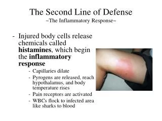
Following injury, trauma or infection of a tissue, a complex series of reactions occur in an effort to prevent ongoing tissue damage, and activate the repair processes. This cumulative homeostatic process is known as inflammation, and the early of reactions are known as the acute phase response (APR). The cells that most commonly initiate the APR are tissue macrophages and blood monocytes. These cells release cytokines such as IL-1 and TNF that control the migration of leukocytes into tissue and orchestrate the inflammatory response. Fever and leuckocytosis are among the most obvious consequences. The biosynthetic activity of the liver is profoundly affected. Acute Phase Response
The pro-inflammatory cytokines IL-1 and TNF act on hepatocytes to greatly increase production of acute phase proteins such as CRP and serum amyloid A protein (Figure 2). These proteins are particularly useful for reflecting inflammation because of the increase of up to 1000 fold from resting levels. There are more moderate increases in the level of other proteins such as ferritin which may affect assessment of iron status in the presence of inflammation. There is a corresponding decrease in the synthesis of some other proteins, most notably albumin which may reach low levels in the presence of sustained inflammation. Acute Phase Response cont…
CRP is more sensitive than ESR to subtle changes in the acute phase response There are distinct ranges of normal and abnormal in CRP reference ranges, without variations for age and gender. CRP is not affected by conditions such as pregnancy, intercurrent drug use, anaemia and plasma protein variations. CRP is a better measure of acute phase response
The phenomenon now recognised as the erythrocyte sedimentation rate was first observed by ancient Greeks. They observed the relationship between the sedimentation of red blood cells and fibrinogen (or “phlegma”),and used it as a means of detecting bad bodily “humors”.16 The test used today was introduced in 1918 by Robin Fahraeus, when he recorded the erythrocytes of pregnant women settled more quickly than the erythrocytes of non pregnant women. ESR
Normally, erythrocytes settle quite slowly, as the gravitational force of the erythrocyte’s mass is counteracted by the buoyant force of the erythrocyte’s volume. In normal circumstances erythrocytes have a net negative charge and therefore they repel each other. Many plasma proteins are positively charged, thereby reducing the repulsive forces and promoting red cell aggregation, which in turn creates a higher mass and therefore the cells settle much more quickly. Fibrinogen and α- and β-globulins are major contributors to the ESR17, and immunoglobulins to a lesser extent. These proteins all have half-lives of days to weeks, and there is a significant lag time between changes at the clinical level and variations in the ESR ESR continued…
If the haematocrit is reduced, as in anaemia for example, the velocity of upward flowing plasma is altered, resulting in the red cells aggregating and therefore falling faster. As a result, in conditions associated with moderate or severe anaemia, the ESR is of limited use. In conditions such as myeloma, the increase in the level of immunoglobulins causes an increase in red cell aggregability, resulting in an elevated ESR. ESR continued….
C-reactive protein (CRP) belongs to the pentraxin family of proteins, which has five identical subunits. It was named because it reacts with the somatic C polysaccharide of Streptococcus pneumonae, and was first discovered in 1930 by Tillet and Francis. Plasma levels begin increasing within 4-6 hours following acute inflammatory stimulus and the half-life of CRP is 5-7 hours, therefore the level of CRP in the blood is regulated solely by its own synthesis. CRP
Anti-infective: CRP can opsonise particles for phagocytosis, and activate complement via the classical pathway. Anti-inflammatory actions:CRP helps in preventing systemic inflammation. CRP aids in the release of neutrophils from blood vessels, while preventing white cell adhesion to vessels in non-inflamed tissues. It is also able to stimulate the release of anti-inflammatory molecules from monocytes. Scavenging actions: Although CRP does not bind to normal cell membranes, it can bind avidly to cells that are undergoing apoptosis or necrosis, possibly because it recognises particular receptors that appear on the surface of dying cells. This in turn binds and activates complement, initiating an inflammatory reaction that attracts neutrophils and monocytes to the site. CRP is reported to have several main functions