Download
1 / 31
320 likes | 570 Views
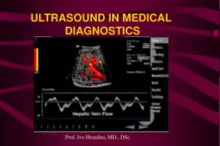
Medical uses of Ultrasound. Bats!. Bats navigate using ultrasound. Bats : Navigating with ultrasound. Bats make high-pitched chirps which are too high for humans to hear. This is called ultrasound Like normal sound, ultrasound echoes off objects

E N D
Bats:Navigating with ultrasound • Bats make high-pitched chirps which are too high for humans to hear. This is called ultrasound • Like normal sound, ultrasound echoes off objects • The bat hears the echoes and works out what caused them • Dolphins also navigate with ultrasound • Submarines use a similar method called sonar • We can also use ultrasound to look inside the body…
Bats:Navigating with ultrasound • If a bat hears an echo 0.01 second after it makes a chirp, how far away is the object? • Clue 1: the speed of sound in air is 330 ms-1 • Clue 2: The speed of sound equals the distance travelled divided by the time taken • Answer: distance = speed x time • Put the numbers in: distance = 330 x 0.01 = 3.3 m • But this is the distance from the bat to the object and back again, so the distance to the object is 1.65m.
Ultrasound imaging:How does it work? • An ultrasound element acts like a bat. • Emit ultrasound and detect echoes • Map out boundary of object
Ultrasound imaging:How does it work? • Now put many elements together to make a probe and create an image
Ultrasound imaging:development of a pregnancy 24 weeks 8 weeks gestation (out of a 40 week pregnancy) 18 weeks
Ultrasound imaging:foetus feet We can process the image in a computer to find the outline of the foot. This is called surface rendering. Here, the foot has been surface rendered This is a 2D ultrasound scan through the foot of a foetus. You can see some of the bones of the foot.
atrium heart valves ventricle Ultrasound imaging:imaging the heart
Doppler effect:change in wavelength with speed • Ultrasound, like normal sound, is a wave. • If a source of sound moves towards the listener, the waves begin to catch up with each other. The wavelength gets shorter and so the frequency gets higher – the sound has a higher pitch. • We use this principle to work out how fast blood cells move. Ultrasound reflects off the blood cells and causes a Doppler shift
The ultrasound probe emits an ultrasound wave • A stationary blood cell reflects the incoming wave with the same wavelength: there is no Doppler shift
The ultrasound probe emits an ultrasound wave • A blood cell moving away from the probe reflects the incoming wave with a longer wavelength • In reality, there is actually two Doppler shifts. The first one occurs between the probe and the moving blood cell (not shown here) and the second one occurs as the red blood cell reflects the ultrasound.
Now, the blood cell moves towards the probe. It reflects the incoming wave with a shorter wavelength
Doppler imaging:combine imaging and Doppler • Use BOTH normal ultrasound imaging and Doppler imaging • Used to image blood flow
Ultrasound imaging:carotid artery • Doppler imaging looks at artery • Get image and trace of blood flow • This is a healthy artery. The flow is smooth and all in the same direction, like water in a large, slow river
Ultrasound imaging:carotid artery • This is also a carotid artery. • The flow is not all in the same direction. It is turbulent, like rapids in a river. • This is usually due to a build-up of fatty deposits in the artery
Ultrasound imaging:4D Doppler ultrasound Ventricles Atria This is a complicated image of the heart of a foetus. It shows the blood moving between the ventricles and the arteries.
Ultrasound:safety • Ultrasound is energy and is absorbed by tissue, causing heating • Question: 2D ultrasound has been used to image the foetus for about 50 years. It is thought to be completely safe and does not cause significant heating • 4D ultrasound is new, requires more energy and therefore generates more heating. We think it is safe. • Should we use it to diagnose foetal illness? • Should we use it to make videos of healthy babies for parents?
Summary: • We can get images of the body by recording echoes of ultrasound • Ultrasound is good at imaging soft tissues • The Doppler effect can be used to detect blood flow
Acknowledgements: • Thanks to GE Healthcare, Prof Jem Hebden and Prof Alf Linney for providing images. • This lesson was developed by Adam Gibson, Jeff Jones, David Sang, Angela Newing, Nicola Hannam and Emily Cook • We have attempted to obtain permission and acknowledge the contributor of every image. If we have inadvertently used images in error, please contact us.