Download

1 / 36
420 likes | 744 Views
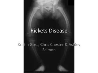
RICKETS IN CHILDREN. DEFINITION : Rickets is consequence of the vitamin D deficit and may occur due to calcium and phosphorus metabolic disorders. Blood analysis shows hypocalcemia and hypophosphatemia. Histology- Failure in mineralisation of the bone and cartilaginous tissues .

E N D
RICKETS IN CHILDREN DEFINITION: Rickets is consequence of the vitamin D deficit and may occur due to calcium and phosphorus metabolic disorders. • Blood analysis shows hypocalcemia and hypophosphatemia. • Histology- Failure in mineralisation of the bone and cartilaginous tissues . • Clinical- manifests as skeletal growth disorder.
Hystory • Rickets ( from Greek word meaning spinal column ) was known since the first years of the • human generation. It is described by Soran Efess (A.D) and by Galen (134-211 A.D). • It is described in detail by a British anatomist and orthopedician, Glisson in 1650. • Incidence: • Rickets is frequently in premature children and the children fed only wheat floor. • In Moldova diagnosis was confirm in 35.5%, X-Ray -21.5% (A.Voloc, M.Garabedian, 1996)
Risk factors • Living in northern latitudes (>30o); • Dark skinned children; • Decreased exposure to sunlight ( polluated geographical areas, humid climate); • Maternal vitamin D deficiency; • Diets low in calcium, phosphorus and vitamin D, e.g. exclusive breast-feeding into late infancy, toddlers on unsupervised “dairy-free” diets; • Macrobiotic, strict vegan diets; • High phytic acid diet, e.g. chapattis; • Prolonged parenteral nutrition in infancy with an inadequate supply of intravenous calcium and phosphate;
Intestinal malabsorption: defective production of 25(OH)D3 – liver disease. Increased metabolism of 25(OH)D3 – enzyme induction by anticonvulsants; Defective production of 1,25(OH)2D3 • Hereditary type I vitamin D-resistant (or dependent) rickets (mutation which abolishes activity of renal hydroxylase); • Familial (X-linked ) hypophosphataemic rickets – renal tubular defect in phosphate transport; • Chronic renal disease; • Fanconi syndrome (renal loss of phosphate) • Target organ resistance to 1,25(OH)2D3- hereditary vitamin D-dependent rickets type II (due to mutations in vitamin D receptor gene).
ETIOLOGY • Rickets is due to partial deficiency, rarely complete deficiency of vitamin D. • Vitamin D exist 2 forms in the human body. • Vitamin D2, exogenous form (calciferol), from ergosterol in the food • Vitamin D3, endogenous form (cholecalciferol or 7-dehydrocholecalciferol from skin), • activated by UV rays of 230-313nm wave length. • Natural alimentation does not supply the daily requirement of 400-500IU of vit.D in a baby. • Breast milk contains 30-50IU/liter, cow’s milk 20-30IU/l, egg yolk contains 20-50IU/10gr. • 80% of the vitamin D is absorbed in the small intestine in the present of normal biliary secretion. • Vitamin D reaches the blood through thoracic duct along with chilomicrons.
Calcium regulation in the blood is as follows: • Vitamin D2 in the food (exogenous) + vitamin D3 (skin, endogenous) =>liver microsomes • =>25(OH) D3 => Mitochondrial kidney tubules membrane activated 3 forms: • 24,25 (OH)2 D3; 1,24,25 (OH)2 D3; 1,25 (OH)2 D3 !!! last more active. • In placental macrophage of pregnancy women are present 1,25(OH)2 D3
FUNCTIONS OF VITAMIN D Intestine: • Increases calcium binding protein • Active transport in the jejunal cells • Phosphorus ions absorption through specific phosphate carrier • Alkaline phosphatase (AP) synthesis • ATP-ase sensibility to calcium ions
Bones • Mineralization of the bone and osteoblast differentiation • Skeletal growth
Kidney • Tubular re-absorption of calcium and phosphorus • Produce 1,25(OH)2D3, the most active form
Muscles • Increases the muscular protein and the ATP in myocytes • Tonicity and the normal contraction of the muscles
Parathyroid glands • Controls parathyroid hormone synthesis • In case of low plasma calcium PTH secretion increases • In response to secretion of PTH increase synthesis of 1,25(OH)2D3 • PTH causes renal losses of phosphate, reducing the bone calcification • Demineralization of bone increase to normal level serum Ca
Other effects of vitamin D • Cellular metabolism: citric acid oxidation • Formation of soluble complex of citrate and Ca in the blood • Skin differentiations in the local treatment of Psoriasis • Pulmonary differentiation (Increases the surfactant in preterm infants) • Immunomodulatory action in autoimmune disorders
Biochemical stages of rickets • Stage 1: Low serum Ca level, normal serum P; normal serum PTH, little raise AP Ca and P tubular re-absorption are normal, no amino acid loss in the urine.
Biochemical stages of rickets Stage 2. Raised PTH in the serum, serum Ca is normalized by bone demineralization. Change in the ratio of Ca : P ( N=2:1), in this stage become 3:1 or 4:1, high serum AP. Raised Ca tubular re-absorption and decrease phosphate tubular re-absorption. As a result => hyper-aminoaciduria. Phosphates are lost in the urine, Ph alkaline. X-ray findings: Osteoporosis and meaphis-epiphesial changes.
Biochemical stages of rickets Stage 3. Severe deficiency of vit.D for a long duration. Laboratory reports: Hypocalcemia, hypophosphatemia, serum elevated of AP, PTH; hyperaminoaciduria, Radiological changes more expressive.
CLASSIFICATION Calcium deficiency rickets can be classified in to 3 grades- I, II, III, Depending on the duration, evolution and the complication: • Grade I, II, III; evolution acute, subacute, recidivant. • Depending on vitamin D insufficiency: • Diet • Infections • Food diversification • Habitual • No prophylaxis • Prophylaxis with low dose • Phenobarbital induced
COMPLICATIONS • Rickets tetany • Convulsions • Respiratory disorders • Cardiac disorders • Skeletal deformation • Frequent illness
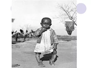
CLINICAL MANIFESTATIONS Rickets may develop in any age of an infant, more frequent at 3-6mo, early in prematures. • The first signs of hypocalcaemia are CNS changes- excitation, restlessness, excessive sweated during sleep and feeding, tremors of the chin and extremities. • Skin and muscle changes- pallor, occipital alopecia, fragile nails and hair, muscular hypotony,motor retardation. • Complications- apnoea, stridor, low calcium level with neuromuscular irritability (tetany). • CNS changes are sometimes interpreted as CNS trauma and the administration of the Phenobarbital which activates the hepatic enzyme may deactivates vit.D and with in 1-2wk of the treatment with Phenobarbital the clinical stage worsens.
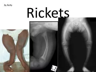
ACUTE SIGNS Have acute and subacute clinical signs • Craniotabes – acute sign of rickets, osteolyses detected by pressing firmly over the occipital or posterior parietal bones, ping-pong ball sensation will be felt. Large anterior fontanella, with hyperflexible borders, cranial deformation with asymmetric occipital flattening.
SUBACUTE SIGNS • Subacute signs are all the following: frontal and temporal bossing • False closure of sutures (increase protein matrix), in the X-ray craniostenosis is absent. • Maxilla in the form of trapezium, abnormal dentition. • Late dental evolution, enamel defects in the temporary and permanent dentition. • Enlargement of costo-chondral junctions-“rickets rosary” • Thorax, sternum deformation, softened lower rib cage at the site of attachment of the diaphragm- Harrison groove.
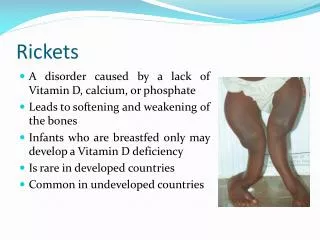
Subacute signs • Spinal column- scoliosis, lordosis, kyphosis. • Pelvis deformity, entrance is narrowed (add to cesarean section in females) • Extremities- palpated wrist expansion from rickets, tibia anterior convexity, bowlegs or knock kness legs. • Deformities of the spine, pelvis and legs result in reduced stature, rachitic dwarfism. • Delayed psychomotor development (heat holding, sitting, standing due to hypotonia).
LABORATORY DATA • Serum calcium level (N=2.2-2.6mmol/l). At the level <2.0mmol/l convulsions sets in. • Phosphorus normal (1.5-1.8mmol/l). Normal ratio of Ca : P= 2:1; in rickets become 3:1; 4:1. • Serum 25(OH)D3 (N=28+2.1ng/ml); and 1,25(OH)2D3(N=0.035+0.003ng/ml) • Serum alkaline phosphatase is elevated >500mmol/l. • Thyrocalcitonin can be appreciated (N=23.6+3.3pM/l) Serum parathyroid hormone (N=598+5.0pM/l) In urine: Aminoaciduria >1.0mg/kg/day • Urinary excretion of 3’5’ cyclic AMP • Decreased calcium excretion (N=50-150mg/24h)
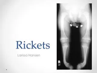
Radiological findings Only in difficult diagnostic cases. • X-ray of the distal ulna and radius: concave (cupping) ends; normally sharply, Fraying rachitic metaphyses and a widened epiphyseal plate. • Osteoporosis of clavicle, costal bones, humerus. • Greenstick fractures. • Thinning of the cortex, diaphysis and the cranial bones.
EVOLUTION The evolution is slow with spontaneous healing at the age of 2-3 years. If treated can be cured in 2-3mo with the normalization of the skeletal and the cellular system. Gibbous, palatal deformity and the narrow pelvis may persist.
DIFFERENTIAL DIAGNOSIS • Osteogenesis imperfecta, chondrodystrophy, congenital diseases- CMV, rubella, syphilis. • Chronic digestive and malabsorption disorders. • Hereditary Fanconi’s disease, phosphorus diabetes, renal tubular acidosis.
PROPHILAXIS IN RICKETS Specific antenatal prophylactic dose administration : 500-1000IU/day of vitamin D3 solution at the 28-th week of pregnancy. The total dose administered is 135000-180000IU. In term infants prophylactic intake of vitamin D2 700IU/d started at 10 days of age during the first 2 years of life; in premature the dose may increase to 1000IU/day.
PROPHILAXIS IN RICKETS WHO recommendation for rickets prophilaxis in a children coming from unfavorable conditions and who have difficult access to hospitals is 200000IU vitamin D2 i/muscular, On the 7day, 2, 4, 6 month- total dose 800000IU. In case of the necessary prolongation 700IU/day till 24mo are given.
SPECIFIC TREATMENT IN RICHETS The treatment is with vitamin D3 depending on the grade. In grade I- 2000-4000IU/day for 4-6weeks, totally 120000-180000IU. In grade II- 4000-6000IU/day for 4-6 weeks, totally 180000-230000IU. In grade III- 8000-12000IU/day for 6-8 weeks, totally 400000-700000IU.
SPECIFIC TREATMENT IN RICHETS • Along with vitamin D, calcium is also administered (40 mg/kg/day for a term baby, • 80 mg/kg/day for a premature baby); also indicate vitamin B&C preparations. • From the 7-th day of the treatment massage can be started. Intramuscular administration • of ATP solution in case of myotonia 1ml/day is preferred.
RICKETS COMPLICATIONS • Rickets tetany in result of low concentration of serum calcium (<2mmol/l), failure of the PTH compensation and muscular irritability occur. • Hypervitaminosis D
Clinical manifestation 1. Manifest tetany: • Spontaneous spasm: flexion at the elbow, extension of 2-5-th digits, extension and adduction of the thumb. • Painful extension and adduction in the tibia tarsal joint. • Rarely contractures in the eyelids and lips muscles. • Laryngeal or bronchial spasm, manifesting as sudden dyspnea, apnea or cyanosis.
Latent tetany: The symptoms are not evident, but they can be performed. Chvostek sign- percussion on the facial nerve leading to contraction of the superior lip, nasal wings, hemi or bilateral facial muscle contraction. Trousseau sign- blood pressure cuff around the mid arm induce carp spasm. Erb sign- <5mA galvanic current induced the nerve impulses. The diagnosis of rickets tetany is based on the clinical manifestation of rickets, low levels of serum calcium, phosphorus, PTH; high serum alkaline phosphatase.
TREATMENT • 1-2% of calcium chloride in milk- 4-6g/day for the first 2 days; after that • 1-3g/day continued for1-2wk. Calcium chloride in more concentrated may cause gastric ulceration. Calcium lactate may be added to milk in 10-12g/d for 10 days. • Oxygen inhalation is indicated in convulsive seizures. Started treatment with vitamin D • 5000-10000IU/d for 6-8weeks, continued calcium intake. When the rickets is healed, the dose of vitamin D should be decrease to the usual prophylactic one.
HYPERVITAMINOSIS D • Symptoms develop in hypersensitivity to vitamin D children or after1-3mo of high doses intakes of vitamin D; they include hypotonia, anorexia, vomiting, irritability, constipation, polydipsia, polyuria, sleep disorder, dehydration. High serum level of acetone, nitrogen and • Ca>2.9mmol/l are found. Increase calcium concentration in urine may provoke incontinence, renal damage and calcification.
Treatment • Preventing calcium rich food, cheese and cow’s milk • Intake mashed fruits and vegetables, juices, hydrating fluids- Ringer solution, water. • Vitamin A, B, E according to age. In severe intoxication administration of Phenobarbital for 2-3 weeks or prednisone 1mg/kg 5-7 days reduces the calcium absorption and increases the calcium excretion. In the case of acidosis 4% sodium hydrocarbonate 5ml/kg is given.
REFERENCES • E.Ciofu, C.Ciofu- Essentials in Pediatrics, 2003. • P.Mogoreanu- Prophylaxis and Treatment of Nutritional Disorders in Children, Kishinau, 2002. • Nelson- Textbook of pediatrics, ed.XVIII, 2008 • Reginald C. Tsang M.B.B.S et al.- Nutrition During Infancy, 1997. • Tom Lissauer, Graham Clayden- Illustrated textbook of Pediatrics, third edition, 2007.