Download
1 / 51
540 likes | 750 Views
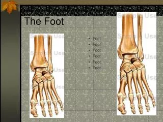
Sole Of The Foot. Dr. Rakesh Kumar Verma Assistant Professor Department of Anatomy KGMU UP Lucknow. SKIN. Thick Nerve Supply. Skin. Skin Thick and hairless.Firmly bound down to the underlying deep fascia by numerous fibrous bands.

E N D
Sole Of The Foot Dr. Rakesh Kumar Verma Assistant Professor Department of Anatomy KGMU UP Lucknow
SKIN Thick Nerve Supply
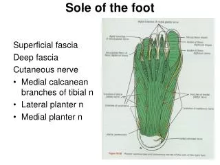
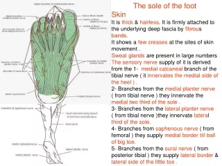
Skin Skin Thick and hairless.Firmly bound down to the underlying deep fascia by numerous fibrous bands. Shows a few flexure creases at the sites of skin movement. Sweat glands are present in large numbers. The sensory nerve supply to the skin of the sole of the foot
Cutaneous Nerves Cutaneus Arteries SUPERFICIAL FASCIA
CUTANEOUS NERVES • Medial calcaneal branch of the tibial nerve • Medial plantar nerve • Lateral plantar nerve • Sural & saphenous nerve
DEEP FASCIA Planter Aponeurosis Deep transverse metatarsal Ligament Fibrous flexor sheath Septae
Plantar aponeurosis • Definition: • Thickened band of deep fascia in the sole of the foot. • Attachment: • Posteriorly: Medial tubercle of calcaneus. • Anteriorly: Divides into 5 slips which pass to the 5 toes. • On each side: Attached to the metatarsal bones by medial and lateral intermuscular septa.
Plantar aponeurosis • Functions: • Protects the underlying nerves and vessels. • Maintains the longitudinal arches of the foot.
MUSCLES FOUR LAYERS FIRST(SUPERFICIAL) SECOND THIRD FOURTH
1st Layer • Three Muscles: • Abductor hallucis • Flexor digitorum brevis • Abductor digiti minimi
SECOND LAYER 1.QUADRATUS PLANTAE/FLEXOR DIGITORUM ACCESSORIOUS 2.LUMBRICALS 3.TENDON OF FLEXOR DIGITORUM LONGUS 4.TENDON OF FLEXOR HALLUCIS LONGUS
2nd Layer • Two Tendons: • Flexor halusis longus • Flexor digitorum longus • Two Muscles: • Quadratus Plantae (Flexor digitorum accessorius) • 4 Lumbricals muscles
3rd Layer • Three Muscles: • Flexor hallucis brevis. • Adductor hallucis • Flexor digiti minimi brevis
FOURTH LAYER 1.DORSAL INTEROSSEI (4) 2.PLANTER INTEROSSEI(3) 3.TENDON OF PERONEUS LONGUS 4.TENDON OF TIBIALIS POSTERIOR
4th Layer • Two Tendons: • Tibialis posterior • Peroneus Longus • Two Muscles: • 3 Planter Interossei • 4 Dorsal Interossei
NERVES MEDIAL PLANTER LATERAL PLANTER
Medial Plantar Nerve It is larger of the two terminal branch of the posterior tibial nerve. Enter the foot midway between the medial malleolus and the medial tubercle of the calcaneus, under cover the flexor retinaculum. Passes forwards deep to the abductor hallucis muscle. Terminate at the bases of the metatarsal bones by dividing into 3 planter digital nerves.
Medial Plantar Nerve • Branches: • Muscular (to four muscles) to: Abductor hallucis. • Flexor digitorum brevis. • Flexor hallucis brevis • First lumbrical muscle • Cutaneous: Planter cutaneous branches: • To the skin of the medial 2/3 of the sole of the foot. • Planter digital nerves • Articular branches: To intertarsal and tarso-metatarsal joints.
Lateral Plantar Nerve It is smaller of the two terminal branches of the posterior tibial nerve. Enters the foot midway between the medial malleolus and the medial tubercle of the calcaneus under cover the flexor retinaculum. Passes forwards and laterally deep to abductor hallusis. Terminate at the base of the 5th metatarsal bone, by dividing into a superficial and a deep branches.
Lateral Plantar Nerve Branches: Muscular : • Flexor digitoum accessorius muscle • Abductor digiti minimi • Flexor digiti minimi brevis • Adductor halucis muscle. • Interossei • 2nd, 3rd & 4th lumbricals. • Cutaneous: • Skin of the lat. 1/3 of the sole • Skin on the lat.side of the planter surface of the little toe and the adjoining sides of the 4th & 5th toes. • The planter digital branches, also, supply the skin on the dorsum of the terminal phalanges of the lateral one and half toes.
Tibial nerve NERVES OF SOLE Lat. Plantar Nerve Med. Plantar Nerve To Flex. Dig. Accessorius To Abd. Digiti Minimi Br. to Abd. Hallucis Br. to Flex. Dig. Brevis Deep Branch Superficial Branch Br. to Flex. Hal. Brevis 1st Lumbrical To Flex. Dig. Min. Brevis & Muscles of 4th interos. space 2nd Lumbrical
Medial Plantar Artery One of the two terminal branches of the posterior tibial artery. Enter the foot midway between the medial malleolus and the medial tubercle of the calcaneus, under cover the flexor retinaculum. Passes forwards deep to the abductor hallucis muscle. Passes b/w the abd.hallucis and flexor digitorum brevis.
Medial Plantar Artery Termination: By anastmosing with the 1st planter metatarsal artery. Branches: Muscular Digital: 3 superficial digital branches these branches end by anastmosing with the first, second and third planter metatarsal arteries.
Lateral Plantar Artery One of the two terminal branches of the posterior tibial artery. At first between the 1st and 2nd layers, then curves medially between the 3rd and 4th layers of the sole. Turns medially with the deep branch of the lateral planter nerve with slight forward convexity to from the plantar arch between the 3rd & 4th layers of muscles.
Lateral Plantar Artery • Branches: • Muscular • Anastomotic branches: b/w arcuate & lateral tarsal arteries of the dorsalis pedis artery. • Posterior perforating arteries: • 3 branches which anastomose with the dorsal metatarsal arteries. • Planter digital artery: to the lateral side of the little toe. • Three planter metatarsal arteries
Transverse section through sole of right foot 3 Prox. Perforating A Dorsalis Pedis A. 4th layer Metatarsal - 1 Metatarsal - 5 3nd layer 2nd layer Deep Br. Of Lat. Plantar art. 1st layer Trunk of Lateral Plantar art.
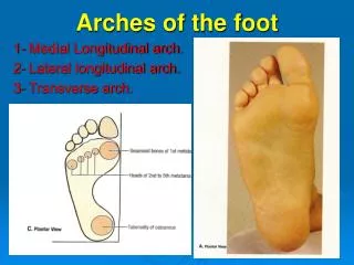
APPLIED ANATOMY ARCHES OF FOOT PAIN MTEATARSALGIA PLANTAR FASCITIS INFECTION INJURY DIABETIC FOOT CALCANEAL SPUR
QUESTION-1 Which dermatome is mainly stimulated in plantar reflex: A) L 4 B) L 5C) S 1 D) S 2
QUESTION-2 All of the following belong to 3rd layer of muscles in sole except: A) Flexor hallucis brevis B) Abductor hallucis C) Adductor hallucis D) Flexor digiti minimi brevis
QUESTION-3 During walking though the flexor digitorum longus contracting strongly, the toes do not buckle because of action of all the following muscles except: A) flexor digitorum accessorious B) Extensor digitorum longus C) Lumbricles D) Interossei
QUESTION-4 Plantar arch mainly formed by medial plantar artery – True/ false