Download
1 / 62
620 likes | 638 Views
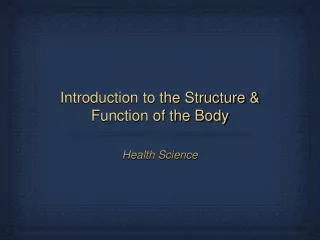
Learn about the levels of organization in the body, the integumentary system, skeletal system, muscular system, and excretory system. Understand the functions and structures of each system, including the importance of maintaining homeostasis.

E N D
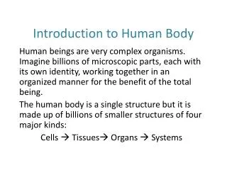
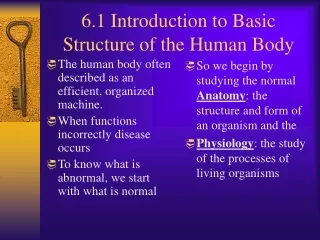
BODY ORGANIZATION • The levels of organization of the body: cells- individual unit tissues- Similar cells that work together to perform a common function. organs- Combination of 2 or more tissues that work together to perform a common function organ system- Group of organs that work together to perform a specific function.
BODY ORGANIZATION Maintaining homeostasis requires: • Body’s organs functioning together. • Temperature regulation (endotherms) • Adjusting metabolism • Detecting and responding to stimuli • Maintaining water and mineral balances
IntegumentarySystem Consists of: Skin, Hair, & Nails
Skin The largest organ in your body. Yes, skin is an organ. Functions of the Skin Protective barrier against pathogens Prevents water loss Offers body protection Regulates body temperature through sweating
Four Tissues of the Integumentary System • Epithelial- covers body surfaces • Connective- provides support and protection • Muscle – body movement • Nerve- forms body’s communication network
3 layers of skin 1. Epidermis: Top layer Constantly makes new skin cells to replace dead ones Contains keratin, which as a waterproof barrier Also contains melanin, a brown pigment that helps protect you from UV rays. (This is why people tan)
3 layers of skin 2. Dermis: The 2nd layer of skin Contains hair follicles (each follicle contains 1 hair) Contains the sebaceous glands which produce an oil called sebum. This lubricates the skin and hair. Contains sweat glands. These release water and some wastes to cool the body and maintain homeostasis
3 layers of skin 3. Subcutaneous tissue: The 3rd layer Composed of fat cells This is used for insulation and an energy supply
SKIN LAYER DIAGRAM EPIDERMIS SEBACEOUS GLAND DERMIS SUBCUTANEOUS TISSUE SWEAT GLAND HAIR FOLLICLE
SKELETAL SYSTEM Functions of the skeleton: • Support the body • Provide protection for the internal organs • Enables movement
SKELETAL SYSTEM There are 206 bones in the skeleton. The skeleton is divided into 2 parts: • axial- includes the skull, spine, ribs, and sternum 2. appendicular- includes shoulders, arms, hips, and legs
SKELETAL SYSTEM Bone is made of hard compact bone surrounding porous bone.
BONE MARROW • RED – makes all blood cells for body (RBC, WBC, & platelets) • YELLOW = stores fat tissue
SKELETAL SYSTEM Early in development, the skeleton is made mostly of hyaline cartilage. Bones hardens as calcium, phosphate and other mineral deposits build up. Osteoblasts make bone tissue. Bones thicken and elongate as development continues.
SKELETAL SYSTEM JOINT = where 2 bones meet. Three types of joints: • Immovable permits little or no movement. ex. skull joined by sutures. • Slightly moveable ex. Spine and ribs • Freely moveable joints (see table 2 p. 854) ex. knee
SKELETAL SYSTEM • Ligament: Connects bone to bone • Tendon: Connects muscle to bone
MUSCULAR SYSTEM Functions Include: • Movement in body • Generate Heat for Body Temperature
MUSCLES Involuntary muscles – not under conscious control. 1. Smooth muscles – line internal organs & blood vessels. a. Function of smooth muscle is to contract. b. Smooth muscle contractions are slow.
MUSCLE 2. Cardiac muscle – heart muscle. Adapted to conduct electrical impulse.
MUSCLE Voluntary muscles – under conscious control skeletal system. 3. Skeletal muscles – attached to the bones & skeletal system. a. Majority of muscles are skeletal b. Contractions are short & strong
MUSCLE Page skeletal muscle structure 1. Skeletal muscle are made up of bundles of muscle fibers. 2. Each muscle fiber is made up of myofibrils.
MUSCLE 3. Myofibrils are made up of smaller proteins filaments. a. Myofibrils are striated or divided into sections called sarcomeres which are the functional units of the muscle
MUSCLE 4. Two types of filaments a. Thick filaments are made up myosin. b. Thin filaments are made up of actin.
MUSCLES Sliding Filament Theory 1. during contractions, actin filaments move towards one another from the pulls of myosin heads
Muscle Fiber • Muscle contractions take a lot of ATP therefore they must have plenty of mitochondria to supply the power.
Human Excretory System • Excretion is the removal of metabolic wastes from the body, including toxic chemicals, excess water, carbon dioxide and salts. • Excretory Organs • Skin • Lungs • Kidneys https://www.youtube.com/watch?v=EhnRhfFLyOg
Nephron Kidney Kidney Ureter Urinary Bladder Urethra Human Urinary System Diagram
Urinary system • The urinary system, consisting of the kidneys, ureters, urinary bladder and urethra, is responsible for eliminating the majority of metabolic wastes from the body • The functional unit of the kidney is the nephron. Each nephron is made of a cup-shaped portion called Bowman’s capsule, tubules and a network of capillaries
Inside the Kidney • Blood pressure within a knot of capillaries (called the glomerulus) increases, causing most of the fluid of the blood to enter Bowman’s capsule • This fluid is called filtrate. • As the filtrate passes through the tubule portion of the nephron, materials needed by the body are reabsorbed and the remainder of the filtrate becomes urine • Proper functioning of the kidney is essential to maintaining homeostatsis in the body
Warm-Up • Write down in correct sequence all the organs (at least 5) through which their food passes as it travels along the digestive tract. Then try to list any glands or organs that are found along the digestive tract, but through which food does not pass.
DIGESTION ANIMATIONS • https://www.youtube.com/watch?v=bo2Ape8JHqA • https://www.youtube.com/watch?v=08VyJOEcDos • https://www.youtube.com/watch?v=Mq9fWzO7Dvw
Cool Facts • Your intestines will grow to at least 25 feet as an adult. Be glad you're not a full-grown horse their coiled-up intestines are 89 feet long! • Food sloshing in the stomach can last 3-4 hours • It takes 3 hours for food to move through the intestine • Food drying up and hanging out in the large intestine can last 18 hours to 2 days! • Americans eat over 2 billion pounds of chocolate a year. • In your lifetime, your digestive system may handle about 50 tons!!
Structures • The GastrointestinaI tract (GI), also called the alimentary canal is the system of organs that take in food, digest it to extract nutrients and expels the waste. These organs are the mouth, pharynx, esophagus, stomach, small intestine and large intestine. • Major Functions: • Ingestion • Digestion • Absorption • Defecation or Excretion
Following the Trail • The process begins in the mouth. • Chewing initiates mechanical breakdown of food and is followed by secretion of saliva, which moistens and lubricates food for swallowing. • Saliva also contains amylases (enzymes), which start the chemical breakdown of carbohydrates. • The swallowing reflex begins in the pharynx and initiates rhythmic waves of smooth muscle contractions called peristalsis. • Peristaltic contractions transport food to the stomach and allow a person to swallow even if he/she are upside down.
Human Digestive System • Digestion is the ability to process food in the body into a form that can be absorbed and used or excreted. • Digestion involves three principle processes: • Mechanical digestion: takes place in the mouth, your teeth chew the food • Chemical digestion: using chemicals to digest/ break down food, this takes place in your mouth and stomach where acid and enzymes mix with the food. • Absorption: pulling nutrients out of the food, occurs in the small intestine
Accessory organs: Organs that help with digestion but are not part of the digestive tract. These organs are the tongue, salivary glands, liver, gall bladder, and pancreas.
Mouth Pharynx Esophagus Liver Stomach Large Intestine Small Intestine Villi Human Digestive System Diagram
Following the Trail II • The stomach contains an extra layer of muscle that aids in mechanically mixing and churning food into a semiliquid form called “chyme.” • Chemical digestion begins with proteins through the action of hydrochloric acid (HCl) and the enzyme, pepsin. • Only water and a few substances, such as aspirin and alcohol, are absorbed by the lining of the stomach.
Following the Trail III • As food enters the small intestine secretions from the liver, gall bladder and pancreas are added .