Download
1 / 48
480 likes | 605 Views
Digestive System. By: Isaías Quezada, Emely Rivera, Carlos Sandoval Per.5. Overview. The digestive system consists of the Alimentary canal: Mouth Pharynx Esophagus Stomach Small intestine Anal canal Accessory structures: Salivary glands Liver Gallbladder Pancreas.

E N D
Digestive System By: Isaías Quezada, Emely Rivera, Carlos Sandoval Per.5
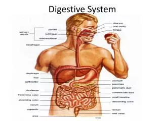
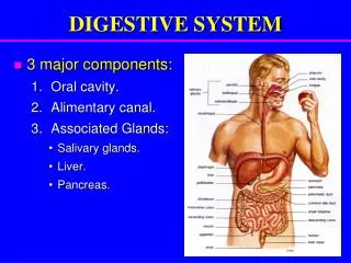
Overview • The digestive system consists of the Alimentary canal: • Mouth • Pharynx • Esophagus • Stomach • Small intestine • Anal canal • Accessory structures: • Salivary glands • Liver • Gallbladder • Pancreas
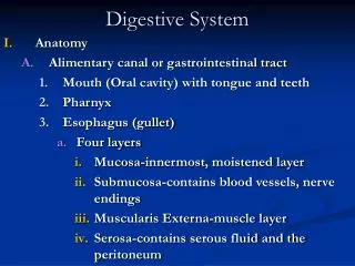
Structure of the Alimentary Canal’s Walls • Its' walls consist of four distinct layers that are developed to different degrees from region to region. Certain regions are specialized for certain functions. These layers are: • Mucosa • Submucosa • Muscular Layer • Serosa
Mucosa • formed of surface epithelium, lamina propria(connective tissue), and a small amount of smooth muscle • Function are protection, secretion, and absorption
Submucosa • Contains loose connective tissue, glands, blood vessels, lymphatic vessels, and nerves • Function is to nourish surrounding tissues and carry away any absorbed materials
Muscular Layer • consists of two coats of smooth muscle tissue arranged in circular and longitudinal groups • Provides movement of the tube and its contents
Serosa • outer covering of the tube and comprised of visceral peritoneum, which is formed of epithelium on the outside and connective tissue beneath • Its function is both protection and lubrication
Types of Movement • There are two main motor functions of the alimentary canal; mixing & propelling
Types of Movement: Mixing • Occurs when smooth muscles in small segments of the tube contract rhythmically • When the stomach is full, waves of muscular contractions move along its wall from one end to the other. • The waves occur every twenty seconds and they mix foods with the digestive juices that the mucosa secretes
Types of Movement: Propelling • a wave-like motion called peristalsis occurs • a ring of contraction appears in the wall of the tube, yet just ahead the muscular wall relaxes • This action begins when food expands the tube
Mouth • First portion of alimentary canal • Receives food • Begins mechanical digestion • Breaks food into smaller pieces • Organ of sensory and speech
Cheeks • Lateral walls of mouth • Consist of outer layers of skin • Pads of subcutaneous fat • Muscles that help with expressions and chewing • Moist inner layers moist, stratified squamious epithelium
Lips • Mobile structures surrounding mouth • Contain skeletal muscle • Judges temperatures and texture of food • Reddish color comes from amount of blood cells • External borders mark boundaries between skin of face and mucous membrane that lines in alimentary canal
Tongue • Muscular organ rest on the bottom of the mouth • Covered by mucous membrane • Connected by midline to the floor by membranous fold, lingual frenulum • Composed of skeletal muscle fibers that run in several directions • Papillae • Root held by hyoid bone • Posterior covered in lingual tonsils
Palate • Roof of oral cavity • Hard anterior , soft posterior • Hard palate formed by palatine processes of maxillary • soft palate forms a muscular arch • Uvula • Muscles here help with swallowing • Palatine tonsils • Pharyngeal tonsils.
Teeth • Hardest structures in body • Not considered part of the skeletal system • Develop in sockets in alveolar process of mandibular and maxillary bones • Permanent teeth come at about six years old • They break food into smaller particles, thus beginning the mechanical process • Helps mix food with saliva
Salivary Glands • Secretes saliva • minor glands are found on mucosa of the mouth • Constantly secreting fluid to keep mouth moist • There are 3 pairs of major glands, parotid, submandibular, sublingual
Secretion • Secretory cells within the glands, Serous, and Mucous cells • Serous cells contain salivary amylase. • Enzymes split starch and glycogen molecules, they then become disaccharides • This begins chemical digestion • Mucous cells secrete mucus • Glands have parasympathetic and sympathetic nerves
Pharynx • Connects nasal and oral cavity with larynx and esophogus Nasopharynx • Located: superior to the soft palate • Communicates with the nasal cavity and provides a passageway for air during breathing Oropharynx • Located: posterior to the mouth • Passageway from the mouth for air moving to and from the nasal cavity
Pharynx (continued) Laryngopharynx • Located: inferior to the oropharynx • Extends from the upper boarder of the….downward to the lower boarder of the cyroid cartilage of the larynx and is a passageway to the esophagus
Esophagus • Straight, collapsible tube that is about 25 cm. long • Provides a passageway for food
Cardiac Sphincter • Remain contracted • Close entrance to the stomach • Prevents regurgitation
Parts of the Stomach: Cardiac Region • A small are near the esophageal opening, or cardia • This is where the contents of the esophagus empty into the stomach
Parts of the Stomach: Fundic Region • Serves as a temporary storage area and sometimes fills with swallowed air • This produces a gastric air bubble which may be used as a landmark on a radiograph of the abdomen
Parts of the Stomach: Body Region • The main portion of the stomach and located between the fundic and pyloric portions • The main chamber for containing food
Parts of the Stomach: Pyloric Region • Funnel-shaped • Shape narrows and becomes the pyloric canal as it approaches the small intestine • At the end, the circular layer of fibers in its muscular wall thickens and forms the muscle Pyloric Sphincter, which acts as a valve that controls gastric emptying
Gastric Secretions • The stomach’s mucous membrane is studded with gastric pits, located at the ends of tubular gastric glands • Their structure and composition of their secretion vary in different parts of the stomach • All gastric glands generally contain 3 types of secretory cells
Secretory Cells • Mucous Cells: found in the necks of the glands near the openings and is responsible for lining the stomach from digestive juices • Chief Cells: also known as peptic cells are a key component of gastric juices • Parietal Cells: also known as oxyntic cells. When the chief cells secrete enzymes and parietal cells release a hydrochloric acid solution, they form the gastric juices
Pancreas • Endocrine Gland/exocrine function • Secretes pancreatic juices
Pancreas (structure) • Located posterior to parietal peritoneum • Pancreatic acinar cells: produce pancreatic cells • Aclai: clusters around tiny tubes which release their secretions • Small tubes connect to pancreatic duct • Pancreatic duct connects with duodenum
Stomach • A J-shaped pouch like organ in the abdominal cavity • It receives food from the esophagus, mixes it with gastric juices, initiates the digestion of proteins, carries on limited absorption, and transports food into small intestine • Divided into four sections; • Cardiac region • Fundic section region • Body regions • Pyloric regions
Liver • Largest gland in the body • Weights 3.2 – 3.7 pounds • Detoxifies blood • Creates bile for stomach • Stores vitamins, iron, simple sugar glucose • Converts ammonia to urea • Very Likely to get disease due to the mass of functions it carries out
Gallbladder • Pear shaped • Stores conventrate bile • Stores any bile that is not used • Cholecystokinin causes the release of bile to small intestine • Cholestoral, bile salts, can create Gallstones
Small Intestine • A tubular organ that extends from the pyloric sphincter to the beginning of the large intestine • Recieves secretions from the pancreas and liver • Completes digestion of the nutrients in chym
Carbohydrates • Begins in the mouth • Parctially digested carbs travel to esophagus and then the stomach • Move to the small intestine where they are broken down by enzymes from the pancreas • Then absorbed into the blood stream • Fiber passes through undigested Macromolecules Lipids • Some digested in mouth and stomach • Mainly in the small intestine • Bile produced in liver is sent to gallbladder • Fatty acids combine with cholesterol and bile • Transported to veins of chest and the blood carries fat to be stored in adipose tissue
Proteins • Digested into amino acids • Begins in stomach with gastric juice • Potent enzymes from pancreas • Continues to the small intestine • Amino acids absorbd into the blood Macromolecules (cont)
Duodenum • Shortest and most fixed portion of the small intestine • C-shaped • Recieves the partly digested food
Jejunum • Greater diameter • Thicker wall • More vascular • More active • Support absorption of carbohydrates and protiens
Ileum • More lymph nodules • Higher bacterial population • Absorb chyme
Large Intestine • 1.5 meters long • Consists of cecum, colon, rectum, and anal canal • Begins in iliac region of pelvis • Joined together with small intestine • Continues across and down the abdominal cavity, and ends at the anus • Takes 16 hours to digest • Transports waste and re-absorption of water before it gets secreted. • Absorbs water and vitamins • Reduces Acidity • Produces antibodies
Cecum • Tube like structure in lower abdominal cavity • Receives undigested food from small intestine • Absorbs fluids and salts that remain after intestinal digestion and absorption • Mixes the contents with mucus • Has a thick layer of mucous membrane • Layer of muscle that makes churning and rubbing movements
Colon • Removes water, salt, and nutrients that form stool • Muscles squeeze the contents through the intestine • Bacteria is found along the walls of the colon • 4 parts, descending, ascending, transverse, and sigmoid • Supported by peritoneum
Rectum • 10 to 12 cm • Dilates towards the anus • Stores feces • Stretch receptors in walls tell when the body needs to defecate • When the rectum storage is full, the pressure pushes the feces to the anus • Body temperature can checked from rectum area
Anal Canal • 3-5 cm • Lubricates feces as it comes from rectum • Has muscular sphincter system that closes lumen • External anal sphincter surrounds anal canal and acts like a clamp. Similar to the puborectalis muscles that covers rectum from behind. • Both of which are voluntarily controlled • Internal anal sphincter relaxes so blood in anal cushions drain, which allows feces to go through