Download
1 / 25
250 likes | 396 Views
Ruptured esophagus after resection of thoraco abdominal aorta aneurysm. 23/9/10. Case presentation. A 50 years old male was transferred from other hospital.

E N D
Ruptured esophagus after resection of thoraco abdominal aorta aneurysm 23/9/10
Case presentation • A 50 years old male was transferred from other hospital. • One day before referal, he was admitted to that hospital because of severe epigastric pain, the doctor suspected peptic perforation. Exploratory laparotomy was done and found retroperitoneal hematoma in upper part of abdoment. The wound was closed and the patient was transferred.
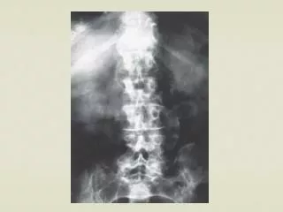
CT angiogram was done after surgery, it showed large thoraco abdominal aortic aneurysm – Crawford type 5 • CAG was done, it was normal.
Left thoracotomy incision was made through 6th intercostal space. The aneurysm extended from mid thoracic to just above celiac artery. The maximal diameter was 10 cm. • Hematoma surrounded the aneurysm. • Left femoral vein was exposed but cannula could not be passed into right atrium. So, the aorta was cross clamped just above the aneurysm. It was opened, clots evacuated, and transected above celiac artery. • A 22 mm dacron graft was anastomosed to distal aorta first , and proximal anastomosis was completed. Aortic clamps were released. The operation and his post operative course was uneventful. Until post operative day 14, he complained of dysphagia and vomitting. He was febrile and chilled.
Chest CT scan showed presence of air in aneurysmal sac surrounding the graft • Emergency left thoracotomy was done. • A large amount of pus and food particles surrounding the graft. • Longituidinal necrosis of lower esophagus about 2 cm long.
Esophagectomy was done with closure of cardia. • Debridement and excision of aneurysmal wall and cleansing of graft. • Open diaphragm and mobilize omental flap. • Cover the graft with omental flap. • Gastrostomy, feeding jejunostomy and cervical esphagectomy were done.
culture • Aortic wall Streptococcal fecalis • Pus Steptococcusfecalis Yeast cell lactobacilli.
Postoperative course after rethoracotomy • Clinically well, he became afebrile and he tolerated jejunostomy feeding fairly. • Two weeks later reopened midline laparotomy was done, while mobilizing the cardia, there was pus from the aneurysmal sac, the omental flap covered the graft well and no necrotic tissue, the space was cleaned and irrgated. • A redivac drain was left in the aneurysmal sac and the abdomen was closed • Post operatively he was well, tolerated jejunostomy feeding, two weeks later the redivac drain was removed and IV antibiotic continued for total two months. • He was discharged to the referring hospital.
Near complete disappearance of perigraft fluid before esophageal bypass
2nd admission • 3 months after previous admission, he was well and wanted to drink and eat by mouth. • Repeated CT chest showed small about of fluid in the aneurysmal sac, much smaller than before. • Right thoracotomy was done and total thoracic esophagectomy was done • One week later, the abdomen was reopened, left side colon was mobilized preserving left colic vessel and the colon was pulled up via retrosternal space and anastomosed to cervical esophagus. Cologastrostomy, colo-colostomy were done.
There was superficial abdominal wound infection. • His post operative course was uneventful otherwise. • Oral diet was resumed on 8th post operative day after contrast study showed no leakage no obstruction.
discussion • Esophageal necrosis after surgery of descending thoracic aortic aneurysm is not rare. • Mechanisms ruptured aneurysm caused pressure on the esophagus surgery excluded aortic branches to esophagus, ischemic necrosis
Prognosis is almost always fatal due to sepsis, mediastinitis prosthetic graft infection extra anatomical bypass – difficult or impossible infection involving suture lines – aortic anastomotic dehiscence -> fatal hemorrhage
Omental graft bactericidal blood supply and white blood cell to combat bacteria cover the prosthetic graft is an important strategy to combat graft infection filling the space surrounding the graft prevent reinfection