Download
1 / 36
360 likes | 649 Views
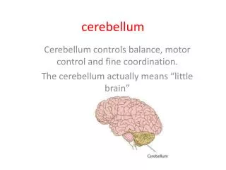
Cerebellum. Won Taek Lee , M.D. Ph.D . Department of Anatomy, Yonsei University College of Medicine. Cerebellum External Configurations. - located in posterior cranial fossa - tentorium cerebelli (cerebrum), 4th ventricle (brain stem) - communicate with other structure via

E N D
Cerebellum Won Taek Lee, M.D. Ph.D. Department of Anatomy, Yonsei University College of Medicine
Cerebellum External Configurations - located in posterior cranial fossa - tentorium cerebelli (cerebrum), 4th ventricle (brain stem) - communicate with other structure via superior, middle, and inferior cerebellar peduncle - longitudinal division Vermis, Paravermal Region, Cerebellar Hemisphere - transverse division Anterior Lobe ------------ primary fissure Posterior Lobe ------------ posterolateral fissure Flocculonodular Lobe
Cerebellum External Configurations Subdivision of Flocculonodular Lobe Nodulus Flocculus Subdivision of Anterior Lobe Vermis Hemisphere Lingula Central Lobule Ala Central Lobule postcentral fissure Culmen Quadriangular Lobule
Cerebellum External Configurations Subdivision of Posterior Lobe Vermis Hemisphere Declive Simple Lobule postcentral fissure FoliumSuperior Semilunar Lobule horizontal fissure Inferior Semilunar Lobule Tuber Gracile Lobule prepyramidal fissure Pyramis Biventer Lobule secondary fissure Uvula Tonsil
Cerebellum Internal Configurations Cerebellar Cortex Molecular Layer Purkinje Cell Layer Granular Layer Corpus Medullare (Medullary Center) Deep Cerebellar Nuclei Fastigial Nuclei Nucleus Interpositus Emboliform Nucleus Globose Nucleus Dentate Nucleus
Deep Nuclei 1. fastigial nucleus 2. globose nucleus 3. emboliform nucleus 4. dentate nucleus
Cerebellum Internal Configurations Cerebellar Cortex I. Molecular Layer Stellate Cell --- taurine (inhibitory) afferent: parallel fiber efferent: Purkinje cell dendrite Basket Cell ---- GABA (inhibitory) afferent: parallel fiber efferent: Purkinje cell soma Parallel Fiber granule cell axon Purkinje Cell Dendrite
Cerebellum Internal Configurations Cerebellar Cortex II. Purkinje Cell Layer Purkinje Cell -- 15,000,000 in number -- GABA (inhibitory) afferent: parallel fiber climbing fiber stellate cell basket cell efferent: deep cortical nuclei Bergman’s glial cell
Cerebellum Internal Configurations Cerebellar Cortex III. Granular Layer Granular Cell -- 50,000,000,000 in number -- glutamic acid (excitatory) afferent: mossy fiber efferent: Purkinje cell dendrite basket cell, stellate cell Golgi cell Golgi Cell -- GABA (inhibitory) afferent: parallel fiber, mossy fiber rosette efferent: granule cell dendrite
1. Purkinje cell 2. granule cell 3. basket cell 4. Golgi cell 5. stellate cell 6. climbing fiber 7. mossy fiber 8. parallel fiber 9. inferior olivary nucleus 10. deep cerebellar nuclei
Cerebellum Internal Configurations Synaptic Glomerulus Afferent terminals on granular layer Mossy Fiber Rosette -- afferent fibers except inferior olivary input -- 2/3 of medullary center Granular Cell Dendrite -- main afferent input Golgi Cell Axon -- synapse on granule cell dendrite -- GABA (inhibitory) - Surrounded by Astrocyte Foot Process
Cerebellum Classifications Classification by Phylogenetic and Ontogenic Development Archicerebellum Paleocerebllum Neocerebellum Classification by Afferent Connection Vestibulocerebellum Spinocerebellum Pontocerebellum Classification by Efferent Connection Vermis Paravermal Region Cerebellar Hemisphere
Archicerebellum (nodulus) Archicerebellum (flocculus) Paleocerebellum Neocerebellum
Spinocerebellum Pontocerebellum Vestibulocerebellum
Cerebellum Connections Afferent Connections (1): 1. Inferior Cerebellar Peduncle Restiform Body Posterior Spinocerebellar Tract Olivocerebellar tract Cuneocerebellar Tract Reticulocerebellar Tract Juxtarestiform Body Vestibulocerebellar Tract Primary Vestyibular Fiber
Cerebellum Connections Afferent Connections (2): 2. Middle Cerebellar Peduncle Pontocerebellar fiber Corticopontocerebellar Fiber Reticulocerebellar Fiber 3. Superior Cerebellar Peduncle Anterior Spinocerebellar Tract Cerulocerebellar fiber Raphecerebellar fiber Rubrocerebellar fiber Hypothalamocerebellar fiber
Cerebellum Connections Efferent Connections : 1. Superior Cerebellar Peduncle Cerebellothalamic fiber - from 3 deep nuclei to VPLo, VLc, CL Cerebellorubral fiber - from nucleus interpositus and dentate nucleus ascending portion of uncinate fasciculus of Russell 2. Inferior Cerebellar Peduncle Fastigiovestibular fiber descending portion of uncinate fasciculus of Russell
Main Connections of the Vestibulocerebellum Vestibular Organ Floculonodular Lobe Vermis VESTIBULAR NUCLEUS vestibulospinal tract MLF FASTIGIAL NUCLEUS lower motor neuron ARCHICEREBELLUM LMN
Main Connections of the Paleocerebellum RED NUCLEUS NUCLEUS INTERPOSITUS rubrospinal tract Inferior Olivry Nucleus ANTERIOR LOBE PARAVERMAL ZONE lower motor neuron PALEOCEREBELLUM SPINAL CORD spinocerebellar tract
Main Connections of the Neocerebellum CEREBRAL CORTEX THALAMUS DENTATE NUCLEUS pyramidal tract Pontine Nucleus POSTERIOR LOBE CEREBELLAR HEMISPHERE lower motor neuron NEOCEREBELLUM LMN
Pyramidal Tract and Associated Circuits upper motor neuron UMN Cerebellum BASAL GANGLIA pyramidal tract lower motor neuron UMN
Cerebellum and Automatic Motor Control Motor Cortex CEREBELLUM Red Nucleus Vestibular Nucleus Reticular Formation Lower Motor Neuron (LMN) Proprioceptors
Cerebellum Connections Corticonuclear Connections A zone ---------- fastigial nucleus medial vestibular nucleus B zone ---------- lateral vestibular nucleus C1, C3 zone --- emboliform nucleus C2 ---------------- globose nucleus D1 ---------------- parvocellular portion of dentate nucleus D2 ---------------- magnocellular portion of dentate nucleus
1. vermis 2. paravermal region 3. cerebella hemisphere 4. nodulus 5. flocculus 6. fastigial nucleus 7. globose nucleus 8. emboliform nucleus 9. dentate nucleus 10. medial vestibular nucleus 11. lateral vestibular nucleus
Cerebellum Connections Olivocerebellar Connections Caudal portion of medial and dorsal accessory olivary nucleus ----------------- vermis of cerebellar cortex (A and B) fastigial nucleus vestibular nucleus Rostral portion of medial and dorsal accessory olivary nucleus ----------------- paravermal region (C1, C2, C3) nucleus interpositus Principal Inferior Olivary Nucleus ----------------- cerebellar hemisphere (D1, D2) dentate nucleus
caudal portion rostral portion Principal Inferior Olivary Nucleus medial and dorsal accessory olivary nucleus
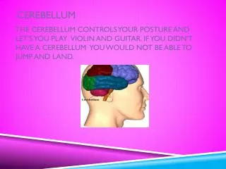
CerebellumFunction Maintenance of Equilibrium - balance, posture, eye movement Coordination of half-automatic movement of walking and posture maintenace - posture, gait Adjustment of Muscle Tone Motor Leaning – Motor Skills Cognitive Function
Motor Skill Pablo Casals
CerebellumClinical Syndromes Ataxia: incoordination of movement - decomposition of movement - dysmetria, past-pointing - dysdiadochokinesia - rebound phenomenon of Holmes - gait ataxia, truncal ataxia, titubation IntentionTremor Hypotonia, Nystagmus Archicerebellar Lesion: medulloblastoma Paleocerebellar Lesion: gait disturbance Neocerebellar Lesion: hypotonia, ataxia, tremor
Posture Gait – Ataxia Tremor
a b c Cerebellar Ataxia Ataxic gait and position: Left cerebellar tumor a. Sways to the right in standing position b. Steady on the right leg c. Unsteady on the left leg d. ataxic gait d
Cerebellar Medulloblastoma Cerebellar tumors on vermis - Truncal Ataxia - Frequent Falling The child in this picture: - would not try to stand unsupported - would not let go of the bed rail if she was stood on the floor.