Download
1 / 20
200 likes | 204 Views
Learn about the endocrine system and its glands, including the pituitary, thyroid, parathyroid, adrenal, and pineal glands. Explore the functions and anatomy of each gland.

E N D
The Endocrine System Dr. Mustafa Saad (2018)
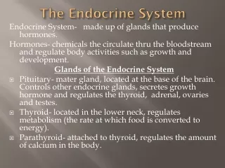
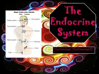
Endocrine Glands A group of secretory cells that release their products, chemical signals called hormones, usually into the circulation. The secretions never pass through ducts • Endocrine glands include: • Pituitary, thyroid, parathyroid, adrenal and pineal glands. • Hypothalamus, thymus, pancreas, ovaries, testes, kidneys, stomach, liver, small intestine, skin, heart, adipose tissue, and placenta also have endocrine function. Fig.1: Endocrine secretion.
The Hypothalamus • Part of the diencephalon of the brain. • Located inferior to the thalamus. • It’s a major link between the nervous and the endocrine systems. • It’s connected to the pituitary gland by blood vessels and nerve fibers. • It secretes a number of hormones that control the secretions of the pituitary gland.
The Pituitary Gland (The Hypophysis) • Located in the hypophyseal fossa of the sphenoid bone. • Considered as the ‘Maestro Gland’ because it releases various hormones that control all the major endocrine glands of the body. Fig.2: The sphenoid bone. • It’s formed of two anatomical and functional parts: • The Adenohypophysis (Anterior Pituitary) • The Neurohypophysis (Posterior Pituitary)
Blood supply of the pituitary • The blood supply derives from two groups of vessels coming off the internal carotid artery: • The inferior hypophyseal arteries provide blood mainly for the neurohypophysis. • The superior hypophyseal arteries form a primary capillary network irrigating the stalk. The capillaries then rejoin to form venules (the hypophyseal portal veins) that branch again as a larger secondary capillary network in the pars distalis. Through this hypophyseal portal system, hormones from the hypothalamus pass to the anterior pituitary gland. • Hormone-rich venous blood leaves the pituitary gland by the anterior and posterior hypophyseal veins
Anterior Pituitary • Has three types of cells: • Acidophils • Basophils • Chromophobes (unstained cells) • The synthesis and release of hormones of the anterior pituitary are controlled by: • Releasing and inhibiting hormones from the hypothalamus. • Feedback regulation. • Hypothalamic hormones made by neurosecretory cells transported by axons to hypophyseal portal system. • Anterior pituitary hormones act on other endocrine glands.
Posterior Pituitary • Does not synthesize hormones. • Releases the following hormones: • Oxytocin • Antidiuretic Hormone (ADH) = Vasopressin • Contains: • Nerve endings • Astrocyte-like cells called Pituicytes. • Cell bodies of neurons in the hypothalamus synthesize these hormone. They’re then transported through axons in the hypothalamo-hypophysial tract to the nerve endings in the posterior pituitary. Here they remain stored. When need arises, they’re released from the nerve endings to pass into blood.
The Thyroid Gland Fig.6: The thyroid gland. • A butterfly-shaped gland located inferior to larynx • Formed of: • Two lobes (right and left) that extend from the thyroid cartilage to the level of 5th tracheal cartilage. They’re related to the: • Common carotid artery and internal jugular vein • External and recurrent laryngeal nerves • Isthmus: a mass of thyroid tissue that connects the two lobes. Located opposite the 2nd, 3rd and 4th tracheal rings. • A Pyramidal lobe may sometimes be present extending superiorly from the isthmus. • The gland is highly vascular. It’s supplied by the superior and inferior thyroid arteries. • It’s covered by a connective tissue capsule.
Pyramidal lobe Fig.7: Anatomy of the thyroid gland.
Histology of the thyroid gland • The thyroid gland consist of numerous spherical structures called the thyroid follicles. • The follicles are lined by epithelial cells (the follicular cells) that range from squamous to columnar according to activity (the more active cells are larger). • Within each follicle there is a lumen into which the thyroid hormones are secreted. • The thyroid gland is unique in that it’s the only endocrine gland in the body that stores its hormones outside the cells. • Between the follicles, there’s another type of cell, called the parafollicular or C-cells, that secrete the hormone calcitonin. Fig.8*: Thyroid follicles and the C-cells.
Fig.9: Histology of the thyroid gland. • The thyroid hormones include: • Tri-iodothyronin (T3) • Tetra-iodothyronin (T4) = thyroxin • Both these hormones affect the body metabolism. • Both these hormones are controlled by TSH from the pituitary gland and by feedback mechanism.
The Parathyroid Gland • Embedded in the posterior aspect of the lobes of the thyroid gland. Each one is surrounded by its own capsule. • They are 4 in number. • Has two types of cells: • Chief (Principal) cells: secrete the Parathyroid Hormone (PTH = parathormone) that regulates level of calcium and phosphate in blood. • Oxyphil cells: Function = Unknown !!?? • Blood calcium level directly controls secretion of both calcitonin and PTH.
The Adrenal (Suprarenal) Glands • Two yellowish glands located on the upper poles of the two kidneys. • The right gland is pyramidal in shape, whereas the left is crescentic in shape. • They’re related to the 12th rib, the diaphragm and the great vessels of the abdomen (aorta and IVC). • Each gland is surrounded by a capsule and composed of two anatomically and functionally distinct region: • Outer yellow cortex: divided into the zona glomerulosa, zona fsciculataand zona reticularis. • Inner brown medulla: a modified sympathetic ganglion that synthesizes and secretes the hormones epinephrine and norepinephrine.