Download
1 / 77
770 likes | 935 Views
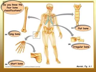
Do you know the four bone classifications?. flat bone. long bone. irregular bone. short bone. Marieb; Fig. 6.1. Can you identify each of these labeled structures in a long bone ?. Marieb; Fig. 6.3. Marieb; Fig. 6.4. Marieb; Fig. 6.4. Can you identify each of these

E N D
Do you know the four bone classifications? flat bone long bone irregular bone short bone Marieb; Fig. 6.1
Can you identify each of these labeled structures in a long bone ? Marieb; Fig. 6.3
Marieb; Fig. 6.4 Can you identify each of these labeled structures in a flat bone ?
Marieb; Fig. 6.4 Can you identify each of these labeled structures in a flat bone ?
In the previous slides, you should have noticed that you labeled compact and spongy bone in both the long and the flat bone. The flat bone is a ‘sandwich’ of spongy bone between compact bone. (And you also did not label an epiphysis or diaphysis in the flat bone.) The cortex of the diaphysis of the long bone is primarily made up of compact bone. However, at its proximal epiphysis, it has a hard outer shell of compact bone, and is filled with spongy bone. We can use these two architectural differences in bone construction (spongy and compact) to identify differences in the function of some bones.
The density of compact bone provides considerable strength and stiffness to our skeletal system. You will see the thickness of compact bone increase down the shafts of long bones such as this femur. These areas must not only respond to high loads imposed down the length of the bone during weight bearing, but must also respond to torsional and bending stresses.
Cancellous bone is also often called spongy bone. This is the type of bone that makes up the interior of most bones (though definitely not the shaft of long bones as you have already seen). Spongy bone is made up of trabeculae. These trabeculae align themselves in the direction of the imposed stress on a bone. The presence of these trabeculae make cancellous bone appear more porous. This makes it lighter in weight than compact bone, but still provides considerable strength. However, it is not as strong as compact bone, so is more easily fractured. Wolff’s Law
These are two vertebrae. The vertebrae are surrounded by an outer layer of cortical bone, and filled with cancellous bone. However, the vertebral body on the right has lost some trabeculae, and become compressed – resulting in a vertebral compression fracture. http://www.uq.edu.au/~anmforwo/bl115/remodelling.html
We are now going to concentrate on the histology of compact bone. This slide looks at the outer edge of this typical bone. Yellow arrow – periosteum Yellow dotted line – ‘cement’ lines surrounding an osteon Blue arrows - Haversian canal Green arrows – osteocytes within their lacunae
The outer part of the compact bone is formed by circumferential lamellae that extend around the entire circumference of the bone. These are laid down under the periosteum AND the endosteum. Sometimes appear more ‘blue’, depending upon staining – comes from presence of chondroitin sulfate (a ‘GAG’ in the ground substance) http://neuromedia.neurobio.ucla.edu/campbell/bone/wp_frame.htm
http://neuromedia.neurobio.ucla.edu/campbell/bone/wp_frame.htmhttp://neuromedia.neurobio.ucla.edu/campbell/bone/wp_frame.htm
Outer edge of compact bone. Green arrows – outer circumferential lamellae White arrows - osteocytes OUTER EDGE
The periosteumis a connective tissue layer on the outer surface of the bone. Periosteum http://www.usask.ca/anatomy/teaching/anat232/Bonejpg/I-18%20Periosteum.jpg
Periosteum http://www.usask.ca/anatomy/teaching/anat232/Bonejpg/I-20%20Periosteum.jpg
At higher magnification, you can see that the periosteumconsists of two layers. • An outer fibrous layer of dense irregular connective tissue, which is quite vascular (remember the dermis?) • An osteogenic layer composed primarily of osteoblasts. This layer is CRUCIAL for the growth of bone.
Outer fibrous periosteum http://www.usask.ca/anatomy/teaching/anat232/Bonejpg/I-21%20Outer%20Fibrous%20Periost.jpg
Sometimes you will see evidence of Sharpey’s fibers at the level of the periosteum. Sharpey’s fibers reflect the sites of the ‘ends’ of tendons or ligaments as their collagen fibers extend deeply into the bone for secure attachment. Sharpey’s Fibers http://www.usask.ca/anatomy/teaching/anat232/Bonejpg/I-21%20Sharpey%27s%20Fibres.jpg
The inner part of the bone is lined by an endosteum. Blue arrows - osteocyte Yellow arrows - endosteum Green arrow - marrow cavity Marieb; Fig. 6.4
So now let’s take a look at the “structural unit” of this compact bone. This structural unit is called either an osteon or an Haversian system. http://neuromedia.neurobio.ucla.edu/campbell/bone/wp_frame.htm Marieb; Fig. 6.5
So what are the names of the three different lamellae in this compact bone? Circumferential lamellae that are located just deep to the periosteum AND to the endosteum Concentric lamellae that make up the osteons or Haversian systems Interstitial system Interstitial lamellae that are usually remnants of old osteons http://www.usask.ca/anatomy/teaching/anat232/Bonejpg/I-19%20Interstit.%20System.jpg
Blue arrows – Haversian canals which house the blood supply to the osteocytes Yellow dotted lines – concentric lamellae White dotted line – interstitial lamellae (remnants of old osteons) Green dotted line – ‘cement’ line that reflects the outer border of the osteon White arrows – osteocytes residing in lacunae
‘Cement’ line http://www.usask.ca/anatomy/teaching/anat232/Bonejpg/I-19%20Cementing%20Line.jpg
Haversian Canal http://www.usask.ca/anatomy/teaching/anat232/Bonejpg/I-18%20Haversian%20Canal.jpg
Blue arrows – outlining osteon or Haversian system Redarrows – Volkmann’s canal (These canals run perpendicular to the Haversian systems or to the long axis of the bone. They connect the blood and nerve supply of the periosteum and endosteum to those in the central Haversian canals.)
http://neuromedia.neurobio.ucla.edu/campbell/bone/wp_frame.htmhttp://neuromedia.neurobio.ucla.edu/campbell/bone/wp_frame.htm
http://neuromedia.neurobio.ucla.edu/campbell/bone/wp_frame.htmhttp://neuromedia.neurobio.ucla.edu/campbell/bone/wp_frame.htm
Blue arrow – osteocyte within its lacunae Yellowarrows – canaliculi What is the role of the canaliculi? Remember that as the osteoblast has finished its work, the osteocyte is now surrounded by several concentric layers of calcified bone. In order to receive nutrients, the osteocytes communicate with other osteocytes via gap junctions by sending cellular projections through these tiny tunnels in the bone called canaliculi.
http://neuromedia.neurobio.ucla.edu/campbell/bone/wp_frame.htmhttp://neuromedia.neurobio.ucla.edu/campbell/bone/wp_frame.htm
Note – most of this information on bone cell types is NOT in your textbook! Two Broad Classifications of Bone Cells Osteoblasts Osteoclasts Bone lining cells - inactive osteoblasts that exist on resting bone surfaces Osteoblasts – active in bone formation Osteocytes – osteoblasts that have become ‘encased’ in osteoid
Osteoclasts • main resorbing cell (engulf collagen, mineral, and osteocytes) • 2. large, multinucleated, and mobile • ruffled border • often reside in depressions called Howship’s lacunae Osteoclast http://www.usask.ca/anatomy/teaching/anat232/Bonejpg/I-22%20Osteoclast%2C%20High%20Mag.jpg
Osteoclast Howship's lacuna carved out by osteoclast http://www.usask.ca/anatomy/teaching/anat232/Bonejpg/II-6%20Osteoclast%2C%20High%20Mag.jpg
Multinucleated osteoclast http://www.usask.ca/anatomy/teaching/anat232/Bonejpg/I-22%20Osteoclast%2C%20Low%20Mag.jpg
http://neuromedia.neurobio.ucla.edu/campbell/bone/wp_frame.htmhttp://neuromedia.neurobio.ucla.edu/campbell/bone/wp_frame.htm
Osteoblasts • main bone forming cell – secretes osteoid (osteoid is nonmineralized matrix) • 2. much smaller than osteoclasts, mononucleated, and usually somewhat cuboidal Osteoblast http://www.usask.ca/anatomy/teaching/anat232/Bonejpg/I-22%20Osteoblasts.jpg
Osteoblast (mononucleated) http://www.usask.ca/anatomy/teaching/anat232/Bonejpg/II-6%20Osteoblast%2C%20High%20Mag.jpg
Blue arrows – osteoblasts Yellow arrows – osteoid (freshly laid down bone that has not yet been mineralized)
Osteocyte • most abundant bone cell type • reside within lacunae • have cytoplasmic extensions that communicate with other osteocytes via gap junctions in canaliculi Osteocyte http://www.usask.ca/anatomy/teaching/anat232/Bonejpg/I-20%20Osteocyte.jpg
Osteocyte in a lacuna http://www.usask.ca/anatomy/teaching/anat232/Bonejpg/I-18%20Osteocyte%20Lacuna.jpg
So, how do these skeletal elements develop? Remember those undifferentiated mesenchymal cells that we talked about in Chapter 4? Well, some of these cells differentiate directly into osteoblastic cells (intramembranous ossification); while others differentiate into chondrocytes which then develop into a hyaline cartilaginous skeleton. Subsequently, this cartilage will be replaced by bone. (endochonral ossification). Marieb; Fig. 4.5
Bone tissue arises by one of two means: (1) intramembranous ossification, or, (2) endochondral ossification. In either case, the original or model tissue is gradually destroyed and replaced with bone tissue. Bony Skeleton Formation Intramembranous Ossification Bone develops directly from fibrous membrane 1. cranial bones of skull 2. diaphysis of clavicle 3. ALSO contributes to the growth of short bones and thickening of long bones Endochondral Ossification Bone develops by replacing hyaline cartilage 1. all bones of skeleton below base of skull EXCEPT clavicles
PROCESSES THAT DESIGN AND REDESIGN BONE Growth Occurs during childhood and the early years of adulthood when the skeleton grows in length and diameter to achieve characteristic external shape Modeling Alters the shape and overall bone architecture during growth and functional adaptation; results when osteoblasts and osteoclasts work independently of one another, and on different surfaces; results in increased bone mass Remodeling Osteoblasts and osteoclasts work closely together to renew old bone of inferior quality and replace bone with micro-fractures; ensures maintenance of calcium homeostasis; generally results in decreased bone mass
So, how does a long bone reach its mature form? Primarily by endochondral ossification. Marieb; Fig. 6.8