Download
1 / 20
220 likes | 397 Views
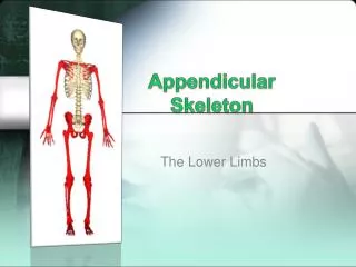
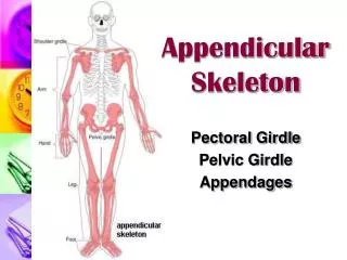
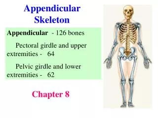
Appendicular Skeleton Continued. Marieb Exercise 11, pgs 151-156. Pelvis and Pelvic Girdle. 2 coxal bones form girdle plus sacrum and coccyx = pelvis anterior articulation at pubic symphysis posterior articulation with sacrum ( sacroilliac ). False Pelvis.

E N D
Appendicular Skeleton Continued Marieb Exercise 11, pgs 151-156
Pelvis and Pelvic Girdle • 2 coxal bones form girdle • plus sacrum and coccyx = pelvis • anterior articulation at pubic symphysis • posterior articulation with sacrum (sacroilliac)
False Pelvis • false (greater): portion superior to pelvic brim or inlet • supports abdominal viscera, but DOES NOT restrict childbirth
True Pelvis • true (lesser): pelvic brim or inlet and inferior • encloses the pelvic cavity • dimensions are critical for childbirth • pelvic inlet: superior most opening • pelvic outlet: inferior most opening
Male vs. Female Pelvis • See Table 11.1 in Marieb for details • Females • wider pelvic inlet and outlet • wider pubic arch
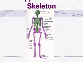
Coxal Bones: 3 fused regions • Ilium, Ischium, & Pubis • Acetabulum (wine cup) • where the 3 bones fuse • recieves the head of the femur • Obturator foramen • hole formed by fusion of pubis and ischium • blood vessels and nerves pass from pelvic cavity into thigh
medial view Ilium • iliac crest • iliac spines mark attachment of muscles and ligaments: • anterior superior iliac spine (ASIS) • anterior inferior iliac spine (AIIS) • Posterior SIS • PIIS • greater sciatic notch • sciatic nerve passes to lower limbs lateral view
medial view ischium • ischial spine • posterior to acetabulum • ischial tuberosity • bear body weight when seated • lesser sciatic notch • blood vessels, nerves and a small muscle pass here lateral view
medial view Pubis lateral view
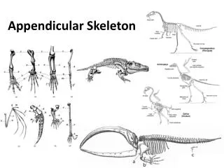
Femur • Greater and Lesser Trochanter • where large tendons attach to the femur • Head • fovea capitis where ligament attaches femur to acetabulum of pelvis • Neck • linea aspera • attachment of hip muscles
Femur • medial/lateral condyles • knee joint • adductor tubercle • adductor magnus muscle • popliteal surface (fossa) • posterior • patellar surface (fossa) • anterior
Patella • A sesamoid bone • enclosed in quadriceps tendon • secures anterior thigh muscle to tibia
Tibia (shin bone) • larger medial leg bone • medial/lateral condyles • knee joint with femur • intercondylar eminence • tibial tuberosity • attachment of patellar ligament
Tibia (shin bone) • medial malleolus • forms inner bulge of ankle • fibular notch • articulates with fibula
Fibula • Lateral smaller bone • lateral malleolus • forms outer bulge of ankle • head • articulates with lateral condyles of tibia
The Foot • 7 tarsals • 5 metatarsals • 14 phalanges
The Foot • 2 largest tarsals support body weight • calcaneus • talus
The Foot • Other 5 Tarsals • Navicular • 1st-3rd cuneiforms • medial, intermediate, lateral • Cuboid
The Foot • Metatarsals (5) • 1st-5th • start at big toe
The Foot • Phalanges (14) • metatarsal 1 • proximal and distal only • metatarsal 2-5 • proximal, middle and distal