Download
1 / 24
440 likes | 1.87k Views
Review of Inflammatory Bowel Disease. Crohn Disease Ulcerative Colitis Pseudomembranous Colitis. Crohn’s Disease. Transmural inflammation involving any part of the GI tract, from mouth to anus. Terminal Ileitius – 80% Ileocolic – 50%

E N D
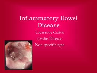
Review of Inflammatory Bowel Disease Crohn Disease Ulcerative Colitis Pseudomembranous Colitis
Crohn’s Disease • Transmural inflammation involving any part of the GI tract, from mouth to anus. • Terminal Ileitius – 80% • Ileocolic – 50% • Colitis – 20% *Differentiate from UC—Crohn’s patients tend to have rectal sparing • Perianal Disease – 30% • Oral and Esophagus – small percentage. • Incidence: Most common 15-40, second peak between 50-80 (bimodal distribution) • Signs/Symptoms: Typical history of prolonged diarrhea with abdominal pain, wt loss and fever +/- gross bleeding. • Characteristics: skip lesions, apthous ulcers, cobblestone appearance (submucosal thickening interspersed with mucosal ulceration) • Treatment: Corticosteroids, aminosalicylates, immune modulators, infliximab (anti-TNF), metronizadole. • Surgery should be avoided if possible since Crohn’s disease is not curable unlike UC. • Complications: Abscess, fistula, obstruction, cancer, perianal disease
Imaging Crohn’s Disease • Small bowel contrast study vs CT • SBFT useful for characterizing length of involvement and areas of stricture • Characteristic Findings • Mucosal nodularity • Narrowed lumen • Ulceration • String sign • Abscesses or fistula • String Sign • Term often applied to the appearance of any marked narrowing of the lumen, but originated as descriptor of reversible narrowing in Crohn disease. • Narrowing caused by incomplete filling as result of irritability/spasms associated with ulceration. String Sign Masselli G. The gastrointestinal string sign. Radiology. 2007 Feb;242(2):632-3.
Ulcerative Colitis • Inflammation confined to mucosal layer of colon that extends from rectum proximally in continuous fashion • Autoimmune process • Incidence: Ages 15-40 or 50-80 (bimodal distribution) • Signs/Symptoms: Rectal bleeding, loose bloody stools, passage of mucus from rectum, abdominal pain • Complications: perforation, stricture, megacolon, cancer • Treatment: • Medical: • Mild/moderate disease—5-ASA, corticosteroids • Severe disease—IV steroids or immunosuppressants for refractory disease • Surgical: Proctocolectomy (curative) • Indications: Failure of medical therapy, increasing risk of cancer with long standing disease, bleeding, perforation • Prognosis: Approximately 1-2% risk of cancer at 10 years, 1%/year thereafter
Barium Enema vs. CT Barium Enema is no longer the test of choice Findings Continuous lesions from rectum proximally with circumferential involvement Imaging Ulcerative Colitis • Lead Pipe Sign • Repeated episodes of mucosal ulceration and marked muscularis hypertrophy results in shortening, narrowing and smoothing out of the normal haustral markings. • “Lead pipe” appearance of colon due to chronic scarring and retraction/loss of haustra Weinstein A et al. A super ‘lead pipe’ colon: radio-pathological correlation of long-standing ulcerative colitis. SA Journal of Radiology;2008 Oct:70-72
Pseudomembranous Colitis • An acute colitis characterized by formation of an adherent inflammatory exudate (pseudomembrane) overlying the site of mucosal injury • Most commonly due to overgrowth of C.difficile, a gram-positive, anaerobic spore forming bacilus • Typically occurs after broad-spectrum antibiotics (especially clindamycin, ampicillin, or cephalosporins) eradicate normal intestinal flora • Signs/Symptoms • Self-limited diarrhea to invasive colitis with megacolon or perforation as possible complications • Diagnosis • Detection of C.diff toxin in stool, proctoscopy or colonoscopy • Treatment • Stop offending antibiotic and give flagyl or vancomycin • Prognosis • High rate of recurrence (20%) despite high response rate to treatment
CT findings • Colonic wall thickening • Target sign • Thickened bowel wall demonstrates three layers that comprise a contrast-enhanced inner and outer layer of high attenuation between which is a layer of decreased attenuation. • Indicates hyperemia in the mucosa and the muscularis propria, serosa, or both with submucosal edema or inflammation • Accordion sign • Alternating edematous haustral folds separated by transverse mucosal ridges filled with oral contrast material, simulating the appearance of an accordion. • Colonic dilatation • Pneumatosis coli or portal venous gas Ahualli J. The target sign: bowel wall. Radiology. 2005 Feb; 234(2):549-550 Macari M et al. The accordion sign at CT: a nonspecific finding in patients with colonic edema. Radiology. 1999 June;211(3):743-746
Case #123 yo male with h/o loose stools and abd pain Diagnosis?
Case #2 • What is the finding? • What is the diagnosis? 3. Which inflammatory bowel disease is this most commonly associated with?
Case #360 year old female presents with abdominal pain, diarrhea, and weight loss. • What is the diagnosis? • Name three complications of this disease.
Case #465 yo male treated with intravenous vancomycin for osteomyelitis of the foot presents with diarrhea and elevated WBC Diagnosis?
Case #5 • What are the findings? • What is the diagnosis?
Case #649 yo female on adjuvant chemotherapy for breast cancer with abd distension and diarrhea • What is the most likely diagnosis? • What are two CT imaging findings that can be seen with this?
Case #123 yo male with h/o loose stools and abd pain Diagnosis? Ulcerative Colitis
What is the finding? • Increased periportal echogenicity • 2. What is the diagnosis? • Sclerosing Cholangitis Case #2 3. Which inflammatory bowel disease is this most commonly associated with? Ulcerative Colitis
Sclerosing Cholangitis • Classic sonographic finding is thickening (increased echogenicity) of intra and extra-hepatic bile ducts • 75% of pts with PSC have inflammatory bowel dz (usually UC)
Sclerosing Cholangitis • Disease is characterized by inflammation, destruction and fibrosis of bile ducts • Increased incidence of bacterial cholangitis and cholangiocarcinoma • Definitive treatment is orthotopic liver transplant • Secondary causes of SC include drugs, prior surgery, multiple opportunistic infections
Case #360 year old female presents with abdominal pain, diarrhea, and weight loss. • What is the diagnosis? • Crohns Disease 2. Name three complications of this disease. Abscess, fistula, obstruction
Case #465 yo male treated with intravenous vancomycin for osteomyelitis of the foot presents with diarrhea and elevated WBC Diagnosis? Pseudomembranous Colitis
Case #5 • What are the findings? • Narrowing of bowel lumen with cobblestoning 2. What is the diagnosis? Crohns Disease
Case #649 yo female on adjuvant chemotherapy for breast cancer with abd distension and diarrhea • What is the most likely diagnosis? • Pseudomembranous Colitis • What are two CT imaging findings that can be seen with this? • Target sign • Accordion sign