Download

1 / 65
690 likes | 890 Views
Inflammatory bowel disease. Name-Manek Jethi Roll number-711. Definitions.

E N D
Inflammatory bowel disease Name-Manek Jethi Roll number-711
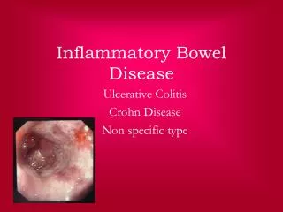
Definitions • Idiopathic inflammatory bowel disease is a set of chronic inflammatory condition resulting from inappropriate and persistent activation of the mucosal immune system, driven by the presence of normal intraluminal flora. • The two disorders known as inflammatory bowel disease are : • Ulcerative colitis (UC)-Ulcerative colitis is an idiopathic chronic inflammatory disease of the colon and rectum, characterized by mucosal inflammation and typically presenting with bloody diarrhea. • Crohn’s disease (CD)-Crohn’s disease is characterized by transmural inflammation of the gut wall and can affect any part of the tubular GI tract
Etiology • Genetic predisposition • HLA-DR1-Crohns disease; HLA-DR2-Ulcerative colitis • NOD2 (CD) • Studies in twins suggest that genetics is a more powerful determinant of disease for CD(67%) than for UC(13%) • Altered/ dysregulated immune response (T-cell response) • Altered response to gut microorganisms • Microbes provide antigens and induce costimulators and cytokines thus triggering immune responses
Pathogenesis • Normal GI protects against foreign pathogens and is unresponsive to normal gut flora • In patients with IBD there are two pathogenic abnormalities- • Strong immune responses against normal flora • Defects in epithelial barrier function • The basis of the abnormalities is still not established which is why they are known as idiopathic
Pathogenesis • In both CD and UC, the prime culprits appear to be CD4+ T-cells, and the lesions are likely caused by T-cells and their products. • Crohndisease appears to be the result of a chronic delayed-type hypersensitivity reaction induced by IFN-y- producing TH1 cells. • The nature of the inflammatory infiltrate, especially the presence of granulomas, is consistent with a TH 1 response. • Animal models suggest that ulcerative colitis is caused by excessive activation of T H2 cells, in the human disease the signature T H 2 cytokine, IL-4, has not been found in the lesions.
Pathophysiology • TH 1- cells appear to induce transmural granulomatous inflammation that resembles CD • TH 2 cells appear to induce superficial mucosal inflammation resembling UC. • The exaggerated T-cell response in both conditions leads to intestinal damage and increased permeability
Clinical features of UC • Hemorrhoids, and fissures, or perirectal abscesses may be present • Iritis, uveitis, episcleritis, and conjunctivitis with ocular involvement • Dermatologic findings with erythema nodosum, pyodermagangrenosum, or aphthous ulceration. • Frequent bowel movements, often with blood in the stool
Lab manifestations of UC • Decreased hematocrit /hemoglobin • Increased erythrocyte sedimentation rate • Leukocytosis and hypoalbuminemia with severe disease. • Two antibodies that can be detected in the serum of IBD patients are- • perinuclearantineutrophil cytoplasmic antibodies (pANCAs) and • anti-Saccharomyces cerevisiaeantibodies (ASCAs).
Complications of UC • Colon adenocarcinoma • extensive mucosal involvement (pancolitis) • family history of carcinoma of the colon. • Perforation • Massive hemorrhage • Toxic megacolon • Defined as a transverse or right colon with a diameter of >6 cm, with loss of haustration in patients with severe attacks of UC.
Clinical manifestations of CD • Abdominal mass and tenderness • Perianal fissure or fistula • Aggressive fistulating disease • Malaise and fever • Abdominal pain • Frequent bowel movements
Lab manifestations of CD • Increased white blood cell count and erythrocyte sedimentation rate • Granulomas are highly characteristic of CD
Complications of CD • Perforation • Abscess formation • Stricture & small bowel obstruction • Nutritional deficiencies • Cancer: small bowel adenocarinoma
Management • The care of a patient with inflammatory bowel disease (IBD) can be either medical or surgical in nature or, in many patients, a combination of both. • The management algorithm is also dependent on whether the diagnosis is Crohn disease or ulcerative colitis. The medical approach for patients with IBD is both symptomatic care (ie, relief of symptoms) and mucosal healing following a stepwise approach to medication, with escalation of the medical regimen until a response is achieved. • The 2 goals of therapy are the achievement of remission (induction) and the prevention of disease flares (maintenance). Note that a top-down approach, with earlier introduction of biologics and immunomodulators, is frequently advocated to forestall complications.
Treatment • The concept of deep mucosal healing, particularly in Crohn disease, is becoming increasingly advocated. • There are several studies, primarily involving anti-TNF agents that have shown that the elimination of inflammation results in a decrease in the rate of surgery, the use of corticosteroids, and the rate of hospitalization • This supports the use of immune-modifying agents (mercaptopurine or azathioprine) or one of the anti-TNF agents earlier in the course of IBD
Treatment procedure • Aminosalicyclates-metronidazole or ciprofloxacin (UC) • Corticosteroids-rapid relief of symptoms and a significant decrease in inflammation • The immune-modifying agents are step III drugs and are used if corticosteroids fail or are required for prolonged periods • In some patients with high-risk disease, a step-down approach with early introduction of stronger agents such as the anti-TNF agents has been advocated to prevent complications and improve patient outcomes
References • Cotran, R. S., & Kumar, V. (2005). Gastrointestinal system.Robbins pathologic basis of disease(7th ed.,). Philadelphia: Saunders. pp. 778-780. • Rowe, W., & Katz, J. (2014, June 2). Inflammatory Bowel Disease Treatment & Management. Inflammatory Bowel Disease Treatment & Management. Retrieved June 8, 2014, from http://emedicine.medscape.com/article/179037-treatment#aw2aab6b6b3
Leukoplakia Name-Manek Jethi Roll number-711
Definition • The term leukoplakia refers to a whitish, well-defined mucosal patch or plaque caused by epidermal thickening or hyperkeratosis • WHO definition-white plaque that cannot be scraped off and cannot be characterized as any other disease • Literal translation-white plates/plaques • Pre-malignant lesion that can give rise to squamous cell carcinoma • Can also occur in the genital area of females. Cause unknown
Appearance of plaques • Localized, sometimes multifocal, even diffuse • Smooth or roughened • Leathery, white, thickened areas • Microscope-hyperkeratosis without underlying epithelial dysplasia to mild to severe dysplasia bordering on carcinoma in situ
Etiology • Plaques are more common in older men • Idiopathic • Tobacco-chewing, smoking, dipped • Chronic alcohol abuse • Ill fitting dentures • HPV • Higher chance in syphilitic patients
Pathogenesis • Hyperplasia of the squamous epithelium • Microscopically-similar to hyperkeratosis of skin • The epithelium is thickened and hyperkeratinized
Pathophysiology • Abnormal epithelial differentiation with increased surface keratinization • May be accompanied by alterations in the epithelial thickness • Underlying molecular changes are uncertain • Marker p53 can indicate pre-malignancy
Clinical features • White or gray plaques • Clinically, Oral Leukoplakia falls into 1 of 2 main groups- • Homogenous oral leukoplakia-uniform white plaques • Speckled or verrucousleukoplakia consists of white flecks or fine nodules on an atrophic erythematous base. These lesions can be regarded as a combination of or a transition between leukoplakia and erythroplasia. High risk of malignancy
Complications • Pre-malignant lesion that can give rise to squamous cell carcinoma • Epithelial changes suggestive of premalignancy include the following: • Nuclear hyperchromatism • Loss of polarity • Increased number of mitotic figures • Nuclear pleomorphism • Altered nuclear-to-cytoplasmic ratio • Deep cell keratinization • Loss of differentiation • Loss of intercellular adherence
Treatment • First step-remove the source of irritation • Quit tobacco/alcohol abuse • If early signs of cancer are detected-surgical removal of plaques • Treatment more successful with early detection and when the lesion is small • Recent study-beta carotene
High yield • A whitish, well-defined mucosal patch or plaque caused by epidermal thickening or hyperkeratosis • Pre-malignant lesion that can give rise to squamous cell carcinoma • Caused by tobacco abuse • Hyperplasia and hyperkeratinization of epithelia
References • Cotran, R. S., & Kumar, V. (2007). The Oral cavity and the Gastrointestinal tract. Robbins pathologic basis of disease (8th ed., ). Philadelphia: Elsevier. • Harris, C. (2013, May 10). Oral Leukoplakia . Oral Leukoplakia. Retrieved June 6, 2014, from http://emedicine.medscape.com/article/853864-overview#showall • Leukoplakia. (2013, July 26). Tests and diagnosis. Retrieved June 6, 2014, from http://www.mayoclinic.org/diseases-conditions/leukoplakia/basics/tests-diagnosis/con-20023802 • Ward, G. E. Leukoplakia. CA: A Cancer Journal for Clinicians, 131-137. Retrieved June 6, 2014, from http://onlinelibrary.wiley.com/store/10.3322/canjclin.3.4.131/asset/131_ftp.pdf;jsessionid=E3E42A916C030AFA83DA2C1BF8917F09.f01t01?v=1&t=hw2nkcmx&s=3c6a14d145d6a3833f91be38668d2cc3fd0bd468
Erythroplakia Name-Manek Jethi Roll number-711
Definition and Etiology • Erythroplakia is a clinical term used to describe a fiery red patch that cannot be clinically or pathologically distinguished as any other definable disease • Red, velvety, possibly eroded area within the oral cavity that usually remains level with or may be slightly depressed than the surrounding mucosa • Atypical lesions • Much higher risk of malignant transformation • Older males at higher risk • Premalignant lesion that can give rise to squamous cell carcinoma
Pathogenesis • Red appearance of the lesion can be attributed to a thin, atrophic epithelium, with a vascular lamina propria lying close to the surface • Secondary infection by Candida albicans • Not confirmed if red surface change in oral erythroleukoplakia is the result of inflammation, dysplasia, or both
Pathophysiology • Rarely epidermal thickening • Superficial erosion of epithelia with dysplasia, carcinoma in situ, or already developed carcinoma • Intense subepithelial inflammatory reaction with vascular dilation leads to red appearance
Clinical features • Asymptomatic, fiery red, well demarcated plaque • Smooth, velvety surface • May be associated with white spots (erythroleukoplakia) • The floor of the mouth, retromolar area, soft palate, and tongue are the most common sites of involvement
Microscopic findings • Abnormal architectural features seen in dysplasia include- • loss of cellular organization or stratification • basal cell layer hyperplasia and loss of polarity • dyskeratosis(single-cell keratinization) • keratin pearls deep within the epithelium • The severity of dysplasia is correlated to severity of the carcinoma in situ
Management/Prevention • Similar to leukoplakia, worse prognosis • Surgical excision of the dysplastic epithelia is the recommended treatment
Differential diagnosis • All diseases with red erythematous patches must be excluded for a diagnosis of erythroplakia • Oral candidiasis • Erythematous candidiasis • Generalized candidial edema • Denture induced stomatitis • Tuberculosis
High yield topics • Erythroplakia is a clinical term used to describe a fiery red patch that cannot be clinically or pathologically distinguished as any other definable disease • Intense subepithelial inflammatory reaction with vascular dilation leads to red appearance • Needs surgical excision for good prognosis
References • Cotran, R. S., & Kumar, V. (2005). Head and Neck. Robbins pathologic basis of disease(7th ed., ). Philadelphia: Saunders. pp. 778-780. • Myers, A. (2013, September 8). Premalignant Conditions of the Oral Cavity . Premalignant Conditions of the Oral Cavity. Retrieved June 6, 2014, from http://emedicine.medscape.com/article/1491418-overview#a30 • Philipsen, H. P. Oral erythroplakia—a review. Oral Oncology, 551-561. Retrieved June 6, 2014, from http://mobi.aslcn1.it/fileadmin/OSRU/Corsi_e_convegni/05._ERITROPLACHIA.pdf
Salivary gland tumors Name-Manek Jethi Roll number-711
Introduction • Salivary gland gives rise to about 30 different neoplasms • A small number of neoplasms makes up about 90% of salivary gland tumors • More common in women • Both benign and malignant tumors about 4-6cm when first diagnosed. Movable on palpation
Pleomorphic adenoma • Most common tumor of salivary glands • 60% of the tumors in the parotid • Mixed tumors • Painless, slow growing tumor, composed of a population of epithelial, myoepethelialand mesenchymalcell components • Definition-A salivary gland tumor that is clinically benign,slow-growing, persistent, characterized by cytomorphologic and architectural diversity, with a tendency to undergo metastatic transformation
Etiology • Unknown • Exposure to radiation • Simian virus • Chromosome 8
Pathogenesis • Roughly 70% of pleomorphic adenomas have cytogenetic alterations that likely play a central role in tumorigenesis , and can be stratified into three groups • those with 8q12 rearrangements, • those with 12q13-15 rearrangements, • those with miscellaneous clonal changes
Pathophysiology • The epithelial elements resembling ductal cells or myoepithelial cells are disposed in duct formations, acini, irregular tubules, strands, or sheets of cells. • The epithelial cells form well-developed ducts lined by cuboidal to columnar cells with an underlying layer of deeply chromatic, small myoepithelial cells. • In most cases, there is no epithelial dysplasia or evident mitotic activity
Clinical features • The palate is the most common intraoral site, followed by the upper lip and buccal mucosa. • They appear as firm, painless, slow-growing swellings and, in the vast majority of cases, do not cause ulceration of the overlying mucosa • Mixed tumors range in size from a few millimeters to several centimeters in diameter • The tumor is typically lobulated and enclosed within a connective tissue pseudocapsule that varies in thickness