Download
1 / 43
870 likes | 4.1k Views
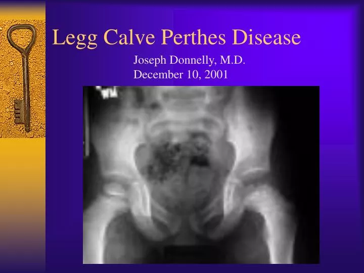
Legg Calve Perthes Disease. Joseph Donnelly, M.D. December 10, 2001. Overview. History Epidemiology/ Etiology Pathogenesis Radiographic stages Presentation/ Exam Imaging Treatment. History. Late 19 th century: “hip infections” that resolved without surgery First described in 1910

E N D
Legg Calve Perthes Disease Joseph Donnelly, M.D. December 10, 2001
Overview • History • Epidemiology/ Etiology • Pathogenesis • Radiographic stages • Presentation/ Exam • Imaging • Treatment
History • Late 19th century: “hip infections” that resolved without surgery • First described in 1910 • Early path studies: cartilaginous islands in the epiphysis
Epidemiology • Disorder of the hip in young children • Usually ages 4-8yo • As early as 2yo, as late as teens • Boys:Girls= 4-5:1 • Bilateral 10-12% • No evidence of inheritance
Etiology • Unknown • Past theories: infection, inflammation, trauma, congenital • Most current theories involve vascular compromise • Sanches 1973: “second infarction theory”
Pathogenesis • Histologic changes described by 1913 • Secondary ossification center= covered by cartilage of 3 zones: • Superficial • Epiphyseal • Thin cartilage zone • Capillaries penetrate thin zone from below
Pathogenesis • Epiphyseal cartilage in LCP disease: • Superficial zone is normal but thickened • Middle zone has 1)areas of extreme hypercellularity in clusters and 2)areas of loose fibrocartilaginous matrix • Superficial and middle layers nourished by synovial fluid • Deep layer relies on blood supply
Pathogenesis • Physeal plate: cleft formation, amorphis debris, blood extravasation • Metaphyseal region: normal bone separated by cartilaginous matrix • Epiphyseal changes can be seen also in greater trochanter, acetabulum
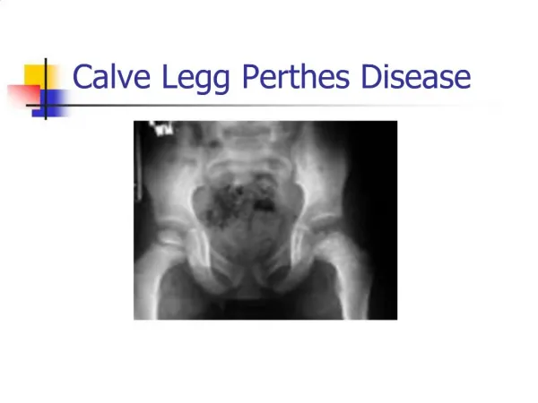
Radiographic Stages • Four Waldenstrom stages: • 1) Initial stage • 2) Fragmentation stage • 3) Reossification stage • 4) Healed stage
Initial Stage • Early radiographic signs: • Failure of femoral ossific nucleus to grow • Widening of medial joint space • “Crescent sign” • Irregular physeal plate • Blurry/ radiolucent metaphysis
Fragmentation Stage • Bony epiphysis begins to fragment • Areas of increased lucency and density • Evidence of repair aspects of disease
Reossification Stage • Normal bone density returns • Alterations in shape of femoral head and neck evident
Healed Stage • Left with residual deformity from disease and repair process • Differs from AVN following Fx or dislocation
Presentation • Often insidious onset of a limp • C/O pain in groin, thigh, knee • 17% relate trauma hx • Can have an acute onset
Physical Exam • Decreased ROM, especially abduction and internal rotation • Trendelenburg test often positive • Adductor contracture • Muscular atrophy of thigh/buttock/calf • Limb length discrepency
Imaging • AP pelvis • Frog leg lateral • Key= view films sequentially over course of dz • Arthrography • MRI role undefined
Differential Diagnosis • Important to rule out infectious etiology (septic arthritis, toxic synovitis) • Others: • Chondrolysis -Neoplasm • JRA -Sickle Cell • Osteomyelitis -Traumatic AVN • Lymphoma -Medication
Radiographic Classifications • Describe extent of epiphyseal disease • Catterall classification= most commonly used • 4 groups based on amount of femoral head involvement • Also presence of sequestrum, metaphyseal rxn, subchondral fx
Lateral Pillar Classification • 3 groups: • A) no lateral pillar involvment • B) >50% lat height intact • C) <50% lat height intact
Salter-Thompson Classification • Simplification of Catterall • Based on status of lateral margin of capital femoral epiphysis • Group A (Catterall I & II equivalent) • Group B (Catterall III & IV equivalent)
Prognosis • 60% of kids do well without tx • AGE is key prognostic factor: • <6yo= good outcome regardless of tx • 6-8yo= not always good results with just containment • >9yo= containment option is questionable, poorer prognosis, significant residual defect
Prognosis • Flat femoral head incongruent with acetabulum= worst prognosis • Do not treat in reossification stage (>15mos)
Non-operative Tx • Improve ROM 1st • Bracing: • Removable abduction orthosis • Pietrie casts • Hips abducted and internally rotated • Wean from brace when improved x-ray healing signs
Non-operative Tx • Check serial radiographs • Q3-4 mos with ROM testing • Continue bracing until: • Lateral column ossifies • Sclerotic areas in epiphysis gone • Cast/brace uninvolved side
Operative Tx • If non-op tx cannot maintain containment • Surgically ideal pt: • 6-9yo • Catterral II-III • Good ROM • <12mos sx • In collapsing phase
Surgical Tx • Surgical options: • Excise lat extruding head portion to stop hinging abduction • Acetabular (innominate) osteotomy to cover head • Varus femoral osteotomy • Arthrodesis
Late Effects of LCP • Coxa magna • Physeal arrest patterns • Irregular head formation • Osteochondritis dessicans