Download
1 / 24
270 likes | 431 Views
Bone & Bone Formation. Written by : RAYAN S. ALBALLAA Histology team, Group A. Edited by: Albara Marwa Histology team, Group A. Components of Bones :. Bone Cells. Calcified Matrix. Periosteum (the outer covering). Endosteum (the inner layer facing the Marrow Cavity).

E N D
Bone & Bone Formation Written by : RAYAN S. ALBALLAA Histology team, Group A Edited by: Albara Marwa Histology team, Group A
Components of Bones : • Bone Cells. • Calcified Matrix. • Periosteum (the outer covering). • Endosteum (the inner layer facing the Marrow Cavity).
Types of Bone Cells : • Osteoprogenitor cells (Forming) • Osteoblasts cells (Immature cells) • Osteocytes (Mature cells) • Osteoclasts
Osteoprogenitor Cells • Arise from MESENCHYMAL STEM CELL. • Differentiate into OSTEOBLASTS. • Found in PERIOSTEUM & ENDOSTEUM.
Osteoblasts : • Derived from OSTEOPROGENITOR cells and can divide. • Have CYTOPLASMIC PROCESSES which are extensions of the cytoplasm. • Basophilic cells on the surface of the bone (in PERIOSTEUM & ENDOSTEUM). • Protein secreting cells. • Secrete the ORGANIC PART OF THE BONE MATRIX.
Osteocytes : • After the osteoblasts secrete the bone matrix, it becomes osteocyte in a small space called the LACUNA. • Osteocytes are mature bone cells with flattened nucleus and cytoplasmic processes. • Can not divide. • Maintain Matrix. • The lacunae (the plural of lacuna) are connected together by small canals called CANALICULI. • Canaliculi contain the cytoplasmic processes of the osteocytes. • GAP JUNCTIONS connect the processes of the osteocytes in the canaliculi.
Osteoclasts : • Multinucleated, motile, and acidophilic cells in the ENDOSTEUM. • Originate by FUSION OF CELLS in the bone marrow. • Secrete Enzymes that digest and remove bone matrix forming cavities and canals (to maintain the Ca++ level in blood). • Have cytoplasmic processes called RUFFLED BORDER.
Bone Matrix : • Bone matrix consists of two components: • organic components : • Type 1 collagen. • Chondroitin sulfate. • inorganic components : • Calcium & phosphorus forming HYDROXYAPATITE CRYSTALS.
Bone Matrix : • In H&E section, the decalcified bone matrix is acidophilic. • It shows the collagen type 1 and the bone cells. • The bundles of collagen in the matrix form parallel layers called bone LAMELLAE.
Periosteum : • It's the outer covering of a bone. • It consists of two layers : • 1/ outer fibrous layer of dense connective tissue attached to a bone by collagen fibers. • 2/ inner cellular layer (Osteogenic layer) of osteoblasts and osteoprogenitor cells. • Function : bone formation and repair.
Endosteum : • it's a layer of cells on the the internal surface of bone facing the marrow cavity. • The cells are the osteoprogenitor cells, osteoblasts, and osteoclasts. • Function : Bone formation and repair.
Types of Bones : Bones exist in two forms : • Compact bone : forms the outer part of all bones in the body. • Cancellous (spongy) bone: forms the inner part of all bones and is more in the epiphysis (the ends of a long bone) than in the diaphysis (the shaft of a long bone).
Compact Bone • The matrix of a compact bone consists of REGULAR lamellae (layers) of calcified type 1 collagen. • The lamellae form parallel cylinders called OSTEONS or HAVERSION SYSTEMS. • Osteons are found deeply in the compact bone.
Compact Bone • The CONCENTRIC lamellae forming the osteons are called OSTEONAL LAMELLAE. • Under the periosteum and endosteum, the lamellae do not form osteons and are called CIRCUMFERENTIAL LAMELLAE.
Canals in a Compact Bone • HARVERSIAN canal in the centre of each osteon contains osteoblasts, osteoclasts, and blood vessels. • VOLKMAN'S canals contain blood vessels and connect the harversion canals of adjacent osteons. • CANALICULI connect the lacunae with Haversion canals for nutrition of the osteocytes.
Cancellous (Spongy) Bones • The lamellae of spongy bone do not form osteons. • The lamellae form INTERCONNECTED TRABECULAE. • (Small pieces of bone).
Cancellous (Spongy) Bones • The lamellae in each Trabecula are parallel to each other. • The Trabeculae are separated by bone marrow spaces lined by endosteum. • In Trabeculae, the canaliculi connect lacunae to bone marrow for nutrition of osteocytes.
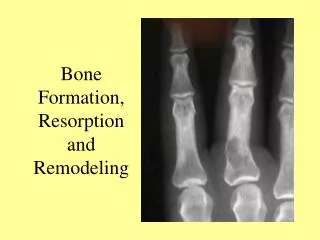
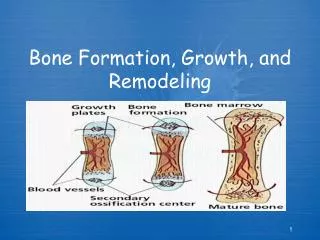
Bone Is Formed by Two Methods : • INTRAMEMBRANOUS OSSIFICATION. • ENDOCHONDRAL OSSIFICATION (INTRACARTILAGENOUS OSSIFICATION).
INTERMEMBRANOUS OSSIFICATION : • In this type of ossification, bone is formed directly in a membrane of MESENCHYMAL cells without the formation of cartilage. (Ex: Flat bones of the skull) • MESENCHYMAL cells differentiate into OSTEOPROGENITOR cells and OSTEOBLASTS which secrete bone matrix and form the PERIOSTEUM. • Calcium salts are deposited in the matrix to form bone. • OSTEOCLASTS remove part of the bone to form MARROW SPACES (Ex: Frontal bone, Maxilla)
ENDOCHONDRAL OSSIFICATION : In this type of ossification, Hyaline Cartilage is formed first and then replaced by bone (Ex: Long bones)
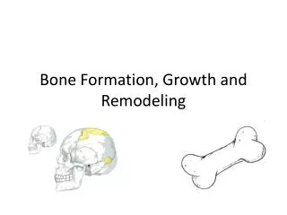
Steps of Endochondral Ossification • MESENCHYMAL cells first form Hyaline cartilage. • Blood vessels enter perichondrium which becomes periosteum and secretes BONE COLLAR on surface of cartilage. • Cartilage in centre degenerates leaving Cavities. • Blood vessels and Osteoblasts from periosteum enter the cavities to form THE PRIMARY OSSIFICATION CENTRE IN THE DIAPHYSIS. • OSTEOBLASTS secrete bone matrix. • OSTEOCLASTS in the ossification centre remove part of the new bone to form the bone marrow cavity.
Epiphyseal Growth Plate of Cartilage • After ossification, a piece of cartilage called EPIPHYSEAL GROWTH PLATE remains between the epiphysis and diaphysis. • Ossification of the growth plate continues up to the age of 20 years. • The growth plate increases the length of bone because its cartilage continues to grow.
Ossification of Epiphyseal Plate Zones of ossification of epiphyseal plate : • Zone of cartilage reserve (resting). • Zone of proliferation of chondrocytes. • Zone of hypertrophy of chondrocytes. • Zone of calcification of cartilage. • Zone of ossification (formation of bone on the calcified cartilage matrix). • Zone of resorption by osteoclasts.