Download
1 / 56
620 likes | 911 Views
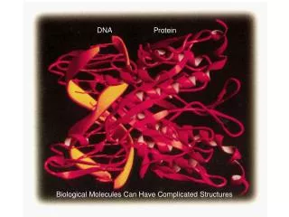
Histology Laboratories Molecules to Systems 2003 Compiled by James D. Jamieson, MD/PhD Thomas L. Lentz, MD. No part of this image collection may be distributed outside of the Yale University Intranet. Acknowledgements. Sources of Micrographs, Diagrams and Figures.

E N D
Histology Laboratories Molecules to Systems 2003 Compiled by James D. Jamieson, MD/PhD Thomas L. Lentz, MD No part of this image collection may be distributed outside of the Yale University Intranet.
Acknowledgements Sources of Micrographs, Diagrams and Figures Alberts, B. et al. Molecular Biology of the Cell. 4th Edition, Garland Science, New York, 2002. Gartner, L. P. and Hiatt, J. L. Color Atlas of Histology, Williams & Wilkins, Baltimore, 1994. Kerr, J. B. Atlas of Functional Histology. Mosby, London, 1999. Kessel, R. G. and Kardon, R. H. Tissues and Organs: a text-atlas of scanning electron microscopy. W. H. Freeman, San Francisco, 1979. Lentz, T. L. Cell Fine Structure. W. B. Saunders, Philadelphia, 1971. Lodish, H. et al. Molecular Cell Biology. W. H. Freeman, New York, 2000. Mizoguti, H. Color Slide Atlas of Histology. Nihon Shashin Shinbunsha, Tokyo. Young, B. and Heath, J. W. Wheater’s Functional Histology. Churchill Livingstone, Edinburgh, 2000. Micrographs taken by George Palade, Marilyn Farquhar, James D. Jamieson, Nicolai Simionescu, Maya Simionescu, David Castle, Thomas L. Lentz. Web Resources http://info.med.yale.edu/webpath/webpath.htm Cushing Library Educational Software/Cell Biology/Several Histology Resources
Some Handy Abbreviations G: glomerulus PCT: proximal convoluted tubule tDL: thin descending limb of Henle tAL: thin ascending limb of Henle TAL: thick ascending limb of Henle DCT: distal convoluted tubule (similar to TAL) CT: connecting/collecting tubule CD: collecting duct MD: macula densa AA: afferent arteriole (in) EA: efferent arteriole (out)
Cortex BV RT G CD Medulla Renal Cortex and Medulla Vasa recta
Renal Corpuscle (Glomerulus, afferent and efferent arterioles, Bowman’s capsule, beginning of proximal convoluted tubule)
Diagram of Renal Corpuscle/Glomerulus
PCT CD G DCT Renal Corpuscles
Bowman’s space G Bowman’s capsule parietal layer visceral layer PCT Renal Corpuscles
MD Vascular pole Bowman’s capsule parietal layer visceral layer G Bowman’s space PCT Urinary pole Renal Corpuscle
Podocytes Bowman’s capsule parietal layer visceral layer PCT Bowman’s space (urinary space) Mesangial cells DCT Renal Corpuscle
Beginning of PCT Urinary Pole of Renal Corpuscle
Bowman’s space Lumen of glomerular capillary Podocyte Foot processes of podocytes Fenestrae Urinary space Urinary space Glomerular BM Fenestrated Capillaries in Glomerulus
Podocyte FS: filtration slits Glomerulus
Filtration slits with diaphragms Capillary space Urinary space Components of Glomerular Filter
Scanning EM of Glomerulus Foot processes Filtration slits
Renal Tubules in Sequence Proximal Convoluted Tubules (PCT): Epithelium: brush border; basal infoldings Loop of Henle: straight portion of PCT (or thick descending limb) (TDL)>thin descending limb (tDL)>loop>thin ascending limb (tAL)>straight portion of DCT (or thick ascending limb) (TAL) Epithelium: Simple squamous epithelium on thin segments; epithelium similar to PCT and DCT on thick segments Distal Convoluted Tubule: Continuous with TAL>macula densa>DCT Epithelium: No brush border, numerous mitochondria Collecting Tubule: Epithelium: cuboidal Collecting Duct: Epithelium: cuboidal
Brush border Proximal Convoluted Tubule
Brush border Proximal Convoluted Tubule
M: mitochondria Mv: microvilli of brush border Proximal Convoluted Tubule
PCT Vasa recta T T CD Thin Walled Tubules of Loop of Henle
VR TAL tL TAL tL CD Relationhship Between CD, TAL, tL and Vasa Recta (VR)
Basolateral infoldings with mitochondria Distal Convoluted Tubule
Basolateral infoldings with mitochondria; few microvilli Distal Convoluted Tubule
Site of urine outflow at renal papilla Renal pelvis Apex of Renal Pyramid
Juxtaglomerular Apparatus and Control of Blood Pressure and Volume
JGA and Control of Blood Pressure
AA: afferent arteriole J: juxtaglomerular cells MD: macula densa DCT: distal conv. tubule JG Apparatus
DT JG Apparatus: Mg, mesangial cells; P, podocyte; JC, juxtaglomerular cells with secretory granules containing renin (arrowheads)
Transitional epithelium Smooth muscle Ureter
Transitional epithelium Relaxed Bladder
Normal Glomerulus and Tubules PAS stain for basement membrane glycoproteins.
Diabetic Kidney PAS stain. What component of the filtration apparatus might you see altered in an EM?
Membranous Glomerulonephritis What structure is the membranous change affecting? The next EM taken at biopsy cinches the diagnosis.