Download
1 / 82
860 likes | 1.3k Views
Osteoporosis. Tracie Wilcox MD Assistant Professor of Medicine University of Chicago PGY 2 Lecture Series. Learning Objectives. Know when to screen for osteoporosis Know how to interpret bone mineral density tests Become familiar with the Frax Bone Treatment Algorithm

E N D
Osteoporosis Tracie Wilcox MD Assistant Professor of Medicine University of Chicago PGY 2 Lecture Series
Learning Objectives • Know when to screen for osteoporosis • Know how to interpret bone mineral density tests • Become familiar with the Frax Bone Treatment Algorithm • Become familiar with the different medications available to treat osteopenia & osteoporosis • Know when and how to screen for secondary causes of osteoporosis
Case 1: • CD is a 60 y.o. WF who presents to her PCP for her annual physical. She has no medical problems, takes no prescription medicines but does take OTC multivitamin and calcium supplements. • Medical History: • postmenopausal since age 55 • HRT from age 55 until 59
Case 1 cont: • Social History: • Rare alcohol • Denies smoking • Walks about 30 minutes 3x per week • Consumes 2 servings of dairy/day + 1000mg Calcium Carbonate • FHX: • Mother suffered hip fracture at age 75
Case 1 cont: • Physical Exam • Ht: 61.5in wt: 152 lbs • Rest of her examination was within normal limits
Case 1 continued • Would you screen her for osteoporosis? • If so, how would you screen?
Background • Approximately 1.5 million fractures annually in US due to osteoporosis • 700,000 vertebral fractures = most common clinical presentation of osteoporosis • 300,000 hip • Increasing risk with age regardless of sex due to combination of decreased bone density and increased risk of falls
Costs • Hip fractures carry 10-20% mortality rate in first year after fracture • Vertebral fractures silent in 2/3 of cases • Estimates of annual costs of osteoporotic fractures $20 billion in US
Risk Factors Major Risk Factors • Previous Fragility fracture • Fragility fracture in first degree relative • Low body weight < 127 lbs (BMI <= 20) • Current smoker (>1PPD) • Postmenopausal status • Age Additional Risk Factors: • >2 drinks/day alcohol • Asian / Caucasian race • Certain medication use and certain clinical conditions – see later
Screening guidelines • USPSTF, NOF, AFP, ISCD all recommend: • Routine screening for women >/= 65 y.o. • For above average risk women (i.e. risk factors other than menopause) begin screening < age 65 • USPTF and AFP recommend starting at age 60 in higher risk women • NOF does not give age for earlier screening
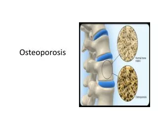
Diagnosis of Osteoporosis • DXA: dual x-ray absorptiometry • Measures bone density at hip and central spine • Can be used to diagnosis osteopenia and osteoporosis • Calculates T and Z scores based upon BMD
T and Z scores • T score: the discrepancy between an individuals BMD and mean peak BMD in a healthy reference population • Z score: the discrepancy between an individuals BMD and an age matched BMD • Can define the relative risk of fracture for age • Score of -2 SD is considered below expected range for age
Other Screening Modalities • Ultrasonography • Indirect assay of bone density • Measures speed or attenuation of ultrasound waves through bone • Can not be used to make diagnosis of osteoporosis as does not measure bone density or bone mineral content directly • Can not be used for monitoring therapy • Benefits: portable and cheap and low levels can predict future fracture risk
Screening Continued • Markers of bone turnover • High values can predict rates of future bone loss • Not used for diagnosis • Used primarily to monitor compliance and efficacy of therapy in patients with continued bone density loss • Check at baseline and then in 6 months after starting therapy • Most commonly check cross-linked N-telopeptide (NTX) and serum carboxy-terminal collagen crosslinks (CTX)
WHO Definitions • Osteoporosis: T-score < -2.5 at lumbar spine, total hip, or femoral neck on bone mineral density scan • Hx of fragility fracture • Osteopenia: -1.0 < T-score <-2.5
Classification • Primary Osteoporosis: describes conditions in which low bone mass is attributed to menopause or aging • Secondary Osteoporosis: describes conditions in which low bone mass is attributed to identifiable factors other than aging and menopause
Secondary Osteoporosis • 20% -30%women thought to have postmenopausal osteoporosis have a secondary cause (1,2) • 2002 Study of 667 postmenopausal women with osteoporosis and low Z scores found 32% had an unrecognized disorder of bone and mineral metabolism(3) 1Harper KD, Weber TJ. Secondary Osteoporosis Diagnostic Considerations. Endocrinology and Metabolism Clinics 1998;27:326-348 2 Lorraine Fitzpatrick. Secondary causes of osteoporosis. Mayo Clinical Proceedings. 2002. 453-468. 3Tannenbaum C, Clark J, Schwartzman K et. Al. Yield of laboratory testing to identify secondary contributors to osteoporosis in otherwise healthy women. J Clin Endocrinol Metab 87:4431-4437;2002
Secondary Osteoporosis • No clear consensus guidelines on when to workup for secondary causes • Consider checking in: • All premenopausal women with osteoporosis • Men • Postmenopausal women with low Z scores (-2.0 or more) • Women who do not respond adequately to initial therapy
Secondary causes of osteoporosis I) Medications: • Glucocorticoids • Aromatase Inhibitors • Depo-medroxyprogesterone acetate • Unfractionated heparin • GnRH agonist • Anticonvulsants: Phenobarbital, Phenytoin, Carbamezapine • via accelerating vitamin D metabolism • Phenothiazine • Methotrexate • Excess vitamin A intake • Cyclosporine-causes severe trabecular bone loss • Tacrolimus-in animal models has demonstrated bone loss
Secondary causes of osteoporosis cont: • 2) Malnutrition/Malabsorption • Celiac Disease • IBD – vitamin D2,D3 largely absorbed in jejunum • Gastric and bowel resection-calcium absorbed in duodenum • Alcohol- inhibits osteoblasts • Vitamin D deficiency/insufficiency
Vitamin D Deficiency • Role of vitamin D: • Increases calcium absorption in the gut • Suppresses PTH release • Decreases renal calcium and phosp excretion • Generation of bioactive vitamin D • During exposure to UV light cholecalciferol ( vit D3) is produced in the skin • Cholecalciferol and ergocalciferol (vitamin D2) also obtained from diet • Vitamin D2 and D3 converted in liver to 25-OH vitamin D (calcidiol) • 1,25-OHD (calcitrol) formed in kidneys through renal conversion
Vitamin D Deficiency • Inadequate levels result in secondary hyperparathyroidism with resultant increase in bone turnover • 25-OH vitamin D levels are regarded as more reliable measure of vitamin D stores • levels >= 30 ng/ml necessary to maximize intestinal calcium absorption and minimize changes in PTH
Vitamin D Deficiency • Prevalence depends on definition • Deficiency: 25-OHD levels < 20 ng/ml • Insufficiency: 25-OHD levels 20 – 30 ng/ml • High prevalence especially in elderly, northern latitudes, pregnancy, IBD, celiac disease
Increased Risk in Elderly • Average person needs 3000-5000 IU of cholecalciferol (D3) per day • Low levels associated with: • Reduced sunlight exposure • Age related decline in 7-dehydrocholesterol in skin • 70 y.o. person has about 25% the level of young adult • Low dietary intake • Oily fish: salmon, mackerel, sardines (400 IU/3.5 oz) • Egg yolks (20 IU) • Fortified foods (milk 100 IU per 8 oz)
Treatment • Vitamin D preparations • Choleocalciferol ( vitamin D3) • Preferred therapy for supplementation as increases 25-OD levels more efficiently than other preparations • give 50,000 IU Q week for 8 weeks then 800 – 1000 IU if deficient • 800-1000 IU per day for insufficiency • Ergocalciferol ( vitamin D2) • Vitamin D metabolites – ex: calcitrol ( 1-25 OHD) • Monitor 25-OHD level 3 months after starting therapy
Secondary causes of osteoporosis cont: • 3) Endocrine: • Hyperthyroidism • Hyperparathyroidism • Cortical bone loss > trabecular • Forearm fracture rates increase • Surgical resection improves BMD • Cushings • Hyperprolactinemia
Secondary causes of osteoporosis cont: • 4) Genetic: • Glycogen storage • Marfans • Ehlers-Danlos Syndrome • Turners • Hemochromatosis-via pituitary involvement
Secondary causes of osteoporosis cont: • 5) Chronic Disease • Multiple Myeloma • uncoupling of bone resorption • stimulation of osteoclast activity (via IL-1, IL6, TNF) • RA • Chronic Renal Disease • Chronic Liver Disease • Systemic Mastocytosis
Secondary causes of osteoporosis cont: • 6) Other • Immbolization- • Study of health volunteers undergoing 12 weeks of bed rest BMD declined 2.9% in spine and 3.8% in hip (1) • Smoker • Post organ transplant- especially first 3-6 months post transplant. • 18-50% prevalence of vertebral fractures postcardiac transplant (1) Zerwekh JE, Ruml LA, Gottschalk F, Pak CY. The effects of twelve weeks of bed rest on bone histology, biochemical markers of bone turnover, and calcium homeostasis iin eleven normal subjects. J Bone Miner Res. 1998;13:1594-1601.
Work Up of Secondary Causes • Influenced by history and physical exam • If no obvious cause consider the following for initial work up • CBC, CMP, 25-OH vitamin D level and calcium excretion • More extensive testing includes urine cortisol, SPEP, UPEP, screening for celiac disease, TSH, PTH, serum or urine markers of bone turnover
2004 US Surgeon General Report recommends a pyramidal approach to osteoporosis treatment
Prevention/Treatment of Osteoporosis • Calcium and Vitamin D • Weight bearing exercise • Medications: • Bisphosphonates • SERM • PTH analogue • Calcitonin
Calcium Intake • 1000-1200mg QD for pre-menopausal and postmenopausal on anti-resorptive therapy • 1500 mg QD for postmenopausal women not on anti-resorptive therapy
Vitamin D Intake • Multiple metanalyses of studies evaluating the effectiveness of vitamin D supplementation on fracture risk • Most evidence shows benefit of supplementation in addition to calcium • Doses need to be 800IU or more / day to decrease fracture risk • Doses of 400 IU/day has not been shown to be effective • Goal of 800 IU / day for prevention to keep 25-OHD levels > 30 ng/ml
Exercise • 30 min three times per week • Small Improvement of BMD • 1 year study of 40 women between ages 50-70 undergoing high intensity (running) exercise showed increase in femoral neck by 0.9% and hip by 1% • In older women has been shown to decrease risk of hip fracture • likely due to increase in muscle strength
When To Start Medication – NOF recommendations • Begin medication in patients with BMD T scores </- 2.5 at femoral neck, total hip, or spine by DXA • Initiate therapy in patients with hip or vertebral fractures
Previous Guidelines National Osteoporosis Foundation (NOF) • T score < -2.0 without risk factors • T score <-1.5 with risk factors • T score based on hip DXA results American Academy of Clinical Endocrinology (AACE) • T score <-2.5 without risk factors • T score < -1.5 with risk factors
WHO Fracture Risk Assessment Tool - FRAX • Released in 2/08 • Algorithm for assessing the 10 year probability of hip fracture and 10 year probability of major osteoporosis-related fracture • Clinical risk factors in the algorithm validated in 60,000 men and women from 12 prospective, population-based cohorts and confirmed in f/u studies
Frax Calculation Tool • Available at http://www.shef.ac.uk/frax • Clinical risk factors: • Age • Sex • Weight / ht • Previous frx • Parent fractured hip • Current smoker • Current steroid use or previous prednisone 5 mg qd/3 mths • RA • Secondary osteoporosis • Alcohol 3 or more units per day • Femoral neck BMD
Frax Calculation Tool • NOF committee collaborated with WHO to perform a cost-effectiveness analysis to estimate the levels of fracture risk above which it is reasonable to consider treatment
New NOF recommendations • Initiate pharmacotherapy in patients with osteopenia and 10 yr hip fracture probability of 3% or 10 year major osteoporosis related fracture probability of 20% based upon the FRAX model
Treatment options • Antiresorptive agents: • Bisphosphonates – first line therapy • SERM • Calcitonin • Estrogen • Anabolic agents: • PTH analogue
Bisphosphonates • Analogues of naturally occurring pyrophosphate • Bind to hydroxyapatite crystals and accelerate osteoclast turnover • Increases BMD and reduces vertebral and nonvertebral fracture rates
Bisphosphonates Alendronate (fosamax) • 10 mg po Q day, 70 mg po Q week • 44% decrease in new vertebral fracture • 56% decrease in hip fracture over average of 4 years Risedronate (Actonel) 5 mg po q day • 41% decrease in new vertebral fracture over 3 years • 39% decrease in nonvertebral fracture • 35 mg po q week, 75 mg po 2x/month • As effective as daily medication and well tolerated • Lancet 1996;348:1535-41. • JAMA 1998;280:2077-82 • JAMA 1999;282:1344-52