Download
1 / 20
221 likes | 523 Views
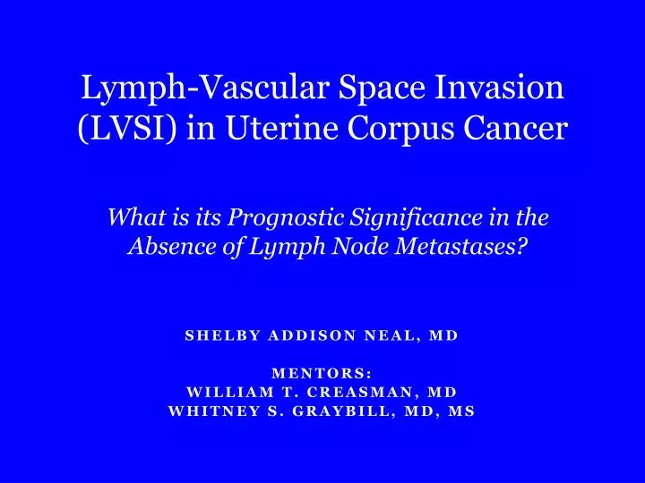
Lymph-Vascular Space Invasion (LVSI) in Uterine Corpus Cancer. What is its P rognostic Significance in the Absence of Lymph Node Metastases?. SHELBY Addison NEAL, MD MENTORS: WILLIAM T. CREASMAN, MD WHITNEY S. GRAYBILL, MD, MS. BACKGROUND.

E N D
Lymph-Vascular Space Invasion (LVSI) in Uterine Corpus Cancer What is its Prognostic Significance in the Absence of Lymph Node Metastases? SHELBY Addison NEAL, MD MENTORS: WILLIAM T. CREASMAN, MD WHITNEY S. GRAYBILL, MD, MS
BACKGROUND Uterine corpus cancer is the most common gynecologic malignancy Surgery is curative in ~80% of cases Adjuvant therapy is appropriate for some patients Rubin and Farber. 1999. Pathology, 3rd edition.
BACKGROUND • Lymph node metsare associated with decreased survival • Lymph-vascular space invasion (LVSI) is an independent risk factor for lymph node metastases1 • OR 6.34, CI 3.45-11.66, P<0.0001 • PPV 33.6% • The presence of LVSI in patients with unstaged endometrial cancer may indicate the need for lymphadenectomy or adjuvant therapy2 • Little data is available regarding LVSI as a prognostic factor in the absence of lymph node metastases 1. Guntupalli et al. 2012. Gynecologic Oncology. 2. Cohn et al. 2002. Gynecologic Oncology.
OBJECTIVE To determine if there is a difference in recurrence-free survival (RFS) or overall survival (OS) between subjects who have LVSI and subjects who do not have LVSI in the setting of negative lymph nodes
HYPOTHESIS In the setting of negative lymph nodes, there is no survival difference between subjects who have LVSI and subjects who do not have LVSI.
STUDY DESIGN • Retrospective chart review of women treated for uterine corpus cancer at MUSC from 1987 to 2012 • Data obtained from charts included the following: • Demographic data • Health history • Surgical pathology • Post-operative clinical course
METHODS Simple regression analysis for the following covariates with recurrence-free and overall survival: Multiple regression analysis incorporating significant covariates • Age • Race • BMI • Parity • Co-morbidities • Histology • Stage • Grade • Lesion size • Depth of invasion • Number of nodes • Adjuvant therapy
METHODS • Model C-index • Measures concordance between predicted and actual survival for each subject • Higher C-index = more accurate model • C-index calculated for 2 different models: • Main model • Model excluding LVSI • Competing risks analysis • Distinguishes between disease-related outcomes and death due to other causes • Outcome of interest = time to recurrence • Competing risk = death due to other causes
COMPETING RISKS ANALYSIS Solid line- LVSI; Dashed line- no LVSI
CONCLUSIONS There is no survival difference between uterine cancer subjects with negative nodes who have LVSI and those who do not Adjuvant therapy for patients with LVSI and negative nodes should not be administered unless otherwise indicated by stage, grade or histology
ACKNOWLEDGEMENTS William Creasman, MD Whitney Graybill, MD, MS Elizabeth Garrett-Mayer, PhD GwenethLazenby, MD Jennifer Pierce, MD, MPH Misty McDowell, MD CatieHaar Virginia McLean Lee Bullard AnquinetteGadson This project was supported by the South Carolina Clinical & Translational Research (SCTR) Institute, with an academic home at the Medical University of South Carolina, through NIH Grant Numbers UL1 RR029882 and UL1 TR000062.