Download

1 / 62
620 likes | 1.4k Views
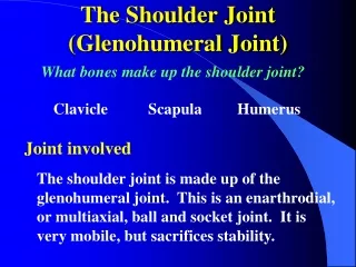
THE SHOULDER JOINT. Ahmad yasin al- zu’bi. Shoulder anatomy. The shoulder joint ( glenohumeral joint) is a ball and socket joint between the scapula and the humerus . It is the major joint connecting the upper limb to the trunk.

E N D
THE SHOULDER JOINT Ahmad yasin al-zu’bi
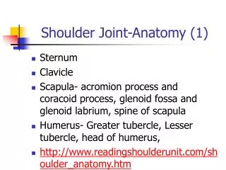
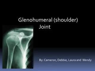
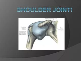
Shoulder anatomy • The shoulder joint (glenohumeral joint) is a ball and socket joint between the scapula and the humerus. It is the major joint connecting the upper limb to the trunk. • It is one of the most mobile joints in the human body, at the cost of joint stability.Joint Capsule and Bursae • The joint capsule is a fibrous sheath which encloses the structures of the joint. • It extends from the anatomical neck of the humerus to the border or ‘rim’ of the glenoid fossa.**glenoid labrum
The synovial membraneproduces synovial fluid to reduce friction between the articular surfaces • synovial bursae:The bursae that are important clinically are: • Subacromial • SubscapularThere are other minor bursae present between the tendons of the muscles around the join
Ligaments • Glenohumeral ligaments (superior, middle and inferior)preventing it from dislocating ? • Coracohumeralligament ? • Transverse humeral ligament? • Coraco–clavicular ligament – composed of the trapezoid and conoidligaments • coracoacromial ligament. Running between the acromion and coracoid process of the scapula it forms thecoraco-acromial arch.??
Movements As a ball and socket synovial joint, there is a wide range of movement permitted: • Extension • Flexion • Abduction-The first 0-15 degrees of abduction is produced by the supraspinatus.-The middle fibres of the deltoid are responsible for the next 15-90 degrees.-Past 90 degrees, the scapula needs to be rotated to achieve abduction – that is carried out by the trapezius and serratus anterior. • Adduction • Internal rotation • External rotation
Factors that contribute to mobility1-Type of joint – ball and socket joint.2-Bony surfaces – shallow glenoid cavity and large humeral head – there is a 1:4 disproportion in surfaces.3-Inherent laxity of the joint capsule. • Factors that contribute to stability:: • Rotator cuff muscles? • Glenoid labrum – a fibrocartilaginous ridge surrounding the glenoid cavity. It deepens the cavity and creates a seal with the head of humerus, reducing the risk of dislocation. • Ligaments–? • Biceps tendon –.?
Neurovasculature • The shoulder joint is supplied by the anterior and posterior circumflex humeralarteries. Branches of the suprascapular artery, a branch of the thyrocervical trunk, also contribute. • innervation is provided by the axillary, suprascapular and lateral pectoral nerves.
Articulating Surfaces • The acromioclavicular joint consists of an articulation between the lateral end of the clavicle and the acromion of the scapula. It has two atypical features: • The articular surfaces of the joint are lined with fibrocartilage (as opposed to hyaline cartilage). • The joint cavity is partially divided by an articular disc – a wedge of fibrocartilage suspended from the upper part of the capsule.
Ligaments • There are three main ligaments that strengthen the acromioclavicular joint. They can be divided into intrinsic and extrinsic ligaments: • Intrinsic: • Acromioclavicular ligament – runs horizontally from the acromion to the lateral clavicle. It covers the joint capsule, reinforcing its superior aspect. • Extrinsic: • Conoid ligament – runs vertically from the coracoid process of the scapula to the conoid tubercle of the clavicle. • Trapezoid ligament – runs from the coracoid process of the scapula to the trapezoid line of the clavicle. • Collectively, the conoid and trapezoid ligaments are known as the coracoclavicular ligament. It is a very strong structure, effectively suspending the weight of the upper limb from the clavicle.
Neurovascular Supply • Vessels • The arterial supply to the joint is via two vessels: • Suprascapular artery – arises from the subclavian artery at the thyrocervical trunk. • Thoraco-acromial artery – arises from the axillary artery. • The veins of the joint follow the major arteries. • Nerves • The acromioclavicular joint is innervated by articular branches of the suprascapular and lateral pectoral nerves. They both arise directly from the brachial plexus.
Movements • The acromioclavicular joint allows a degree of axial rotation and anteroposterior movement. • As no muscles act directly on the joint, all movement is passive, and is initiated by movement at other joints (such as the scapulothoracic joint)??
history • Pain from the shoulder or its surrounding---referred painpain analysis?? • Stiffness may be progressive and severe – so much so as to merit the term ‘frozen shoulder’. • Deformity >1-muscle wasting2-prominence of the acromioclavicular joint 3-winging of the scapula • Instability? • Loss of function?
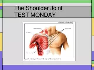
examination • Exposure?Because shoulder and neck symptoms are often felt in the same areas, examination of the shoulder must include a full examination of the neck, and vice versa. • Look■ Skin■ Shape>from front. > from behindAsk the patient to flex the elbow: the appearance of an unnatural bulge over the deltoid muscle is the classic sign of a ruptured biceps tendon.■ Position: if the arm is held persistently internally rotated, think of posterior dislocation of the shoulder.
FeelThe supraspinatus tendon lies just below the anterior edge of the acromion. Tenderness and crepitus can often be localized to a particular structure. • Moveactive passive • abduction(1) difficult to initiate; (2) diminished in range; (3) altered in rhythmIf movement is painful, the arc of pain must be noted>>pain in the mid range of abduction suggests??>>pain at the end of abduction is often?? • Flexion and extension • Adduction • Rotation
Passive movements: these can be deceptive ?? • Power>deltoid>serratus anterior (long thoracic nerve) (winged scapula)>Pectoralis major • Other systems: the cervical spine should be examined as it is a common source of referred pain. If the shoulder feels unstable, look for generalized joint laxity. If weakness is the main complaint, a neurological examination is needed.
Imaging • x-ray • At least two x-ray views should be obtained:1- an anteroposterior in the plane of the glenoid2- an axillary projection with the arm in abduction to show the relationship of the humeral head to the glenoid. • Look for evidence of subluxation, or dislocation, joint space narrowing, bone erosion and calcification in the soft tissues. Special views can show the acromioclavicular joint and the subacromial space
Magnetic resonance imaging (MRI) is useful to identify osteonecrosis of the humeral head, or a bone tumour. It can also identify labral tears and rotator cuff tears although the accuracy for these latter two is enhanced by combining the scan with arthrography. Computed tomography (CT) arthrography is an alternative • Ultrasound is a simple and accurate test for identifying rotator cuff tears and calcific tendinitis. It can also be useful in guiding injections or aspirating calcific deposits in the rotator cuff tears. • Arthroscopy an endoscope that is inserted into the joint through a small incision. is a minimally invasive surgical procedure is useful for diagnosing and treating subacromial impingement, intra-articular lesions, detachment of the glenoid labrum and rotator cuff tears.
Disorders of the rotator cuff • rotator cuff syndrome • calcific tendinitis • adhesive capsulitis.
rotator cuff syndrome • – which comprises several conditions with distinct clinical features and natural history: • subacute tendinitis (painful arc syndrome); • chronic tendinitis (impingement syndrome); • rotator cuff tears
Pathology • The differing clinical pictures stem from three basic pathological processes: • degeneration,with advancing age, the cuff degenerates; minute tears develop, and there may be scarring, fibrocartilaginous metaplasia or calcium depositionThe common site is the ‘critical zone’ of the supraspinatus??is an area approximately 8-15 mm from the insertion of the rotator cuff tendons onto the greater tubercle of the humeral head, mainly within the supraspinatus tendon. This is a watershed zone between the anterior and posterior circumflex humeral, thoracoacromial, and suprahumeral arteries. • TraumaTrauma and impingement: the supraspinatus tendon is liable to injury if it contracts against firm resistance; this may occur when lifting a weight, or when using the arm to save oneself from falling. This is much more likely if the cuff is already degenerate. An insidious type of trauma is attrition of the cuff due to impingement against the coracoacromial arch during abduction. The long head of biceps also may be abraded to the point of rupture. Small tears of the cuff or the long head of biceps are found at autopsy in almost everyone aged over 60 years. • vascular reaction.Vascular reaction: in an attempt to repair a torn tendon or to revascularize a degenerate area, new blood vessels grow in and calcium deposits are resorbed. This vascular reaction may cause congestion and pain
These three pathological processes can be summed up as ‘wear’, ‘tear’ and ‘repair’. • young patient • The older patient • Thus acute tendinitis (which affects younger patients) is intensely painful but rapidly better; chronic tendinitis (a middle group) is only moderately painful but takes many months to recover and may be complicated by partial tears; and a complete tear (which generally occurs in the elderly) becomes painless soon after injury, but never mends.
Symptoms and signs • pain and/or weakness during certain movements of the shoulder. • the patient may know precisely which movements now re-ignite the pain and which to avoid, providing a valuable clue to its origin. • ‘Rotator cuff’ Tenderness is felt at the anterior edge of the acromion • Pain and tenderness directly in front along the deltopectoral boundary could be associated with the biceps tendon. • Localized pain over the top of the shoulder is more likely to be due to acromioclavicular pathology • pain at the back along the scapular border may come from the cervical spine
All these sites should be inspected for muscle wasting, carefully palpated for local tenderness and constantly compared with the opposite shoulder. • If there is weakness with some movements but not with others, then one must rule out a partial or complete tendon rupture; here again, as with pain, localization to a specific site is the key to diagnosis.
clinical examination should include provocative tests to determine the source of the patient’s symptoms: • The painful arc : on active abduction scapulohumeral rhythm is disturbed and pain is aggravated as the arm traverses an arc between 60 and 120 degrees. Repeating the movement with the arm in full external rotation may be much easier for the patient and relatively painless.
Neer’s impingement sign:manoeuvreif positive : 1- Supraspinatus tendinitis 2-long head tendinitis • If the previous manoeuvre is positive, it may be repeated after injecting 10 mL of 1% lidocaine (local anesthetic)into the subacromial space; if the pain is abolished (or significantly reduced), this will help to confirm the diagnosis.
Subacute tendinitis • The patient develops anterior shoulder pain after vigorous or unaccustomed activity, e.g. competitive swimming • Point tenderness is most easily elicited by palpating this spot with the arm held in extension, thus placing the supraspinatus tendon in an exposed position anterior to the acromion process; with the arm held in flexion the tenderness disappears.
Chronic tendinitis • The patient, usually aged between 40 and 50 years, gives a history of recurrent attacks of subacute tendinitis, the pain settling down with rest or antiinflammatory treatment, only to recur when more demanding activities are resumed. • Characteristically pain is worse at night • slight stiffness of the shoulder may restrict even simple activities such as hair grooming or dressing • A disturbing feature is coarse crepitation when the shoulder is passively rotated; this may signify a partial tear or marked fibrosis of the cuff.
Rotator cuff tears • The most advanced stage of the disorder is progressive fibrosis and disruption of the cuff, resulting in either a partial or full thickness tear. • The patient is usually aged over 45 years and gives a history of refractory shoulder pain with increasing stiffness and weakness • Chronic tendinitis vs partial tear >> not easily detected >> imaging • Partial tear vs complete tear >> local anaesthetic • A full thickness tear may follow a long period of chronic tendinitis, but occasionally it occurs spontaneously after a sprain or jerking injury of the shoulder. There is sudden pain and the patient is unable to abduct the arm. • If some weeks have elapsed since the injury the two types are more easily differentiated With a complete tear, pain has by then subsided and the clinical picture is unmistakable: active abduction is impossible and attempting it produces a characteristic shrug; but passive abduction is full and once the arm has been lifted above a right angle the patient can keep it up by using the deltoid (the ‘abduction paradox’); when they lower it sideways it suddenly drops (the ‘drop arm sign’). • In long-standing cases of partial or complete rupture, secondary osteoarthritis of the shoulder may supervene and movements are then severely restricted.
Imaging • X-rays are usually normal in the early stages of the cuff dysfunction, but with chronic tendinitis there may be erosion, sclerosis or cyst formation at the site of cuff insertion on the greater tuberosity, or over-growth of the anterior edge of the acromion, thinning of the acromion process and upward displacement of the humeral head. Osteoarthritis of the acromioclavicular joint is common in older patients and in late cases the glenohumeral joint also may show features of osteoarthritis. Sometimes there is calcification of the supraspinatus but this is coincidental and not the cause of the pain • MRI can effectively show cuff pathology but it should be remembered that up to one-third of asymptomatic individuals also have abnormalities of the rotator cuff on MRI. Changes on MRI always need to be correlated with the clinical examination. • Ultrasonography has comparable accuracy to MRI for identifying and measuring the size of full thickness and partial thickness rotator cuff tears, but is not as accurate in predicting the reparability of the tendons
Conservative treatment • Uncomplicated impingement syndrome (or tendinitis) is often self-limiting • Patients should be taught ways of avoiding the ‘impingement position’ • Physiotherapy, including ultrasound and active exercises • A short course of non-steroidal anti-inflammatory medication sometimes brings relief • If all these methods fail, and before disability becomes marked, the patient should be given one or two injections of depot corticosteroids into the subacromial space. • Healing is slow, and a hasty return to full activity will often precipitate further attacks of tendinitis
Surgical treatment for impingement • indications : • If symptoms do not subside after 3 months of conservative treatment • if they recur persistently after each period of treatment • an operation is considered preferable to prolonged and repeated treatment with anti-inflammatory drugs and local corticosteroids. • The indication is more pressing if there are signs of a partial rotator cuff tear and in particular if there is good clinical evidence of a full thickness tear in a younger patient.
acromioplasty • The object is to decompress the rotator cuff by removing the structures pressing upon it – the coracoacromial ligament, the anterior part of the acromion process and osteophytes at the acromioclavicular joint. • This can be achieved by:1- open surgery2-arthroscopicy>>Advantages of the arthroscopy include less soft-tissue damage, faster rehabilitation and a better cosmetic appearance. • Massive tears which cannot be approximated might be treated by decompression and debridement alone, or with tendon transfer or tendon grafts.
Frozen shoulder: • Adhesive capsulitis (also known as frozen shoulder) is a painful and disabling disorder of unclear cause (idiopathic) in which the shoulder capsule, the connective tissue surrounding the glenohumeraljoint of the shoulder, becomes inflamed and stiff, greatly restricting motion and causing chronic pain. Pain is usually constant, worse at night, and with cold weather. • No known cause (idiopathic) • People who suffer from adhesive capsulitis usually experience severe pain and sleep deprivation for prolonged periods due to pain that gets worse when lying still and restricte movement. • The condition tends to be self-limiting and usually resolves over time without surgery. Most people regain about 90% of shoulder motion over time. • it is NOT caused by secondary causes, so it differs from shoulder stiffness • first movement to be lost in frozen shoulder is → external rotation, while first movement to be lost in shoulder stiffness is → abduction • X-rays are normal. The main role of an x-ray is to exclude other causes of pain and stiffness.
phases of frozen shoulder: • from 0-6 months: “freezing” or painful stage ○ pain increases gradually, range of motion decreases gradually • ● from 6-12 months: “frozen” or adhesive stage ○ the pain is the maximum, range of motion is the minimum • from 1- 1.5 year : "thawing" or recovery stage ○ pain decreases until it disappears, range of motion increases until it becomes near normal.
Differential diagnosis • After any severe shoulder injury, stiffness (without much pain) may persist for some months. It is maximal at the start and gradually lessens, unlike the pattern of a frozen shoulder. • Disuse stiffness If the arm is nursed overcautiously (e.g. following a wrist fracture) the shoulder may stiffen. Again, the characteristic pain pattern of a frozen shoulder is absent. • Regional pain syndrome This condition, formerly known as reflex sympathetic dystrophy, may follow acute trauma; it is also seen in patients with myocardial infarction or a stroke. The features can be similar to those of a frozen shoulder • Arthritis Both rheumatoid arthritis and osteoarthritis can affect the shoulder, and either of these can develop bilaterally. The diagnosis is usually obvious on x-ray
treatment • Pain and inflammation can be controlled with analgesics and NSAIDs. • A physician may also perform manipulation under anesthesia, which breaks up the adhesions and scar tissue in the joint to help restore some range of motion.
Calcific tendinitis: acute and chronic • Acute calcific tendinitis • Calcium hydroxyapatite crystals are deposited in the supraspinatus tendon, probably due to fibrocartilaginous metaplasia from local ischaemia. • Calcification alone is probably not painful; symptoms, when they occur, are due to the vascular reaction which produces swelling and tension in the tendon. • Resorption of the calcific material is rapid and it may soften or disappear entirely within a few weeks. • The condition affects 30–50 year olds. Aching, sometimes following over-use, develops and increases in severity within hours, • During the acute stage the arm is held immobile; the joint is usually too tender to permit palpation or movement. • After a few days, pain subsides and the shoulder gradually returns to normal.
Treatment • X-rays>> Calcification just above the greater tuberosity is always present. As pain subsides, the dense blotch lightens and may then disappear. • the patient is given a short course of nonsteroidal anti-inflammatory medication. • If pain is more intense then corticosteroid is injected into the subacromial space. • Extracorporeal shockwave • ‘barbotage’..drainage • For patients with disabling or recurrent symptoms unresponsive to conservative measures, relief can be obtained by an operation to remove the calcific material.
Chronic calcification • Asymptomatic calcification • appears as an incidental finding in shoulder x-rays. • When it is seen in association with the impingement syndrome, it is tempting to attribute the symptoms to the only obvious abnormality – supraspinatus calcification. However, the connection is spurious and treatment should be directed at the impingement lesion rather than the calcification.
Shoulder Dislocation: • ● Separation of the humerus from the scapula at the glenohumeral joint. • Most commonly dislocated large joint (mostly anteriorly) • ● Less common in children as their epiphyseal plate is weaker and tends to fracture before dislocating • ● More common in elderly as the collagen fibres have fewer cross links → weaker capsule tendons / ligaments. • The main mechanism of dislocation is throwing position (,which is abduction, external rotation and extension). Handball sport is the most common cause that causes dislocation in western countries, but here the most common cause is الطوش