Download
1 / 2
20 likes | 121 Views
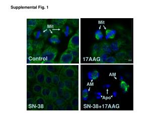
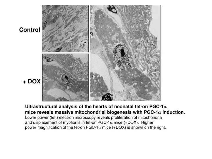
Control. + DOX. Ultrastructural analysis of the hearts of neonatal tet-on PGC-1 a mice reveals massive mitochondrial biogenesis with PGC-1 a induction. Lower power (left) electron microscopy reveals proliferation of mitochondria

E N D
Control + DOX Ultrastructural analysis of the hearts of neonatal tet-on PGC-1a mice reveals massive mitochondrial biogenesis with PGC-1a induction. Lower power (left) electron microscopy reveals proliferation of mitochondria and displacement of myofibrils in tet-on PGC-1a mice (+DOX). Higher power magnification of the tet-on PGC-1a mice (+DOX) is shown on the right.
Baseline Tet-on -4 wks Tet-off-4 wks Cardiomyopathy and myocardial ultrastructural changes in the tet-on PGC-1a mice are reversible. Echocardiography was performed at baseline, after 4 weeks of doxycycline administration (PGC-1a induction) and after 4 weeks post doxyclycline removal (recovery). M-Mode images of LV function (top) and doppler measurements of transmitral flow (bottom) are shown.