Download
1 / 24
260 likes | 544 Views
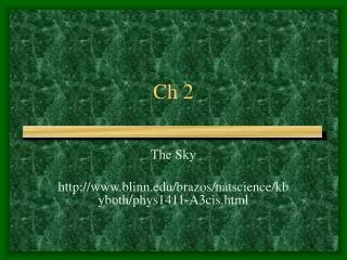
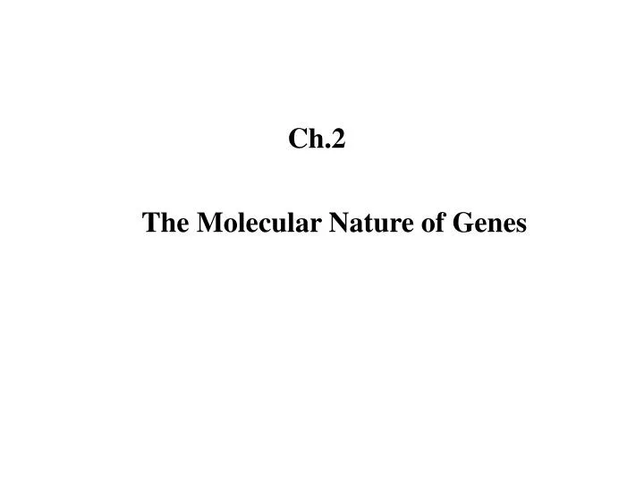
Ch.2. The Molecular Nature of Genes. DNA 의 구조 연구. History of Molecular Genetics. ( S 형 ). ( R 형 ). 유전물질의 발견 ; Miescher (1869) nuclein ; 염색질 (DNA + 단백질 ) 핵산 (nucleic acid) 뉴클레오티드 : 염기 + 당 + 인산 형질전환 (transformation) : Griffith (1928) 박테리아의 형질전환 ;

E N D
Ch.2 The Molecular Nature of Genes
DNA의 구조 연구 History of Molecular Genetics
( S 형 ) ( R 형 ) • 유전물질의 발견 ;Miescher (1869) • nuclein ; 염색질 (DNA + 단백질) • 핵산 (nucleic acid) • 뉴클레오티드 : 염기 + 당 + 인산 • 형질전환 (transformation) : Griffith (1928) • 박테리아의 형질전환 ; • 죽은 S형의 형질전환 물질이 R형에 전달됨 Variants of Streptococcus pneumoniae The nature of genetic material
DNA ; The transforming Material • 형질전환물질은 DNA임을 증명 : Avery 등 (1944) • DNA 가수분해효소에 의해 형질전환 능력 상실 • 물리-화학적 분석 • 초원심 분리법 ; 고분자량의 물질 • 전기영동법 ; 높은 이동성 • UV-흡수분광도법 ; 260 nm의 파장을 흡수 • 화학적 원소분석법 ; N:P의 비율이 낮음 (1.67) • 박테리오파아지의 감염 : Hershey & Chase (1952) • DNA 염기는 같은 비율로 존재하지 않음 : Chargaff (1950) • DNA 이중나선 구조 모델 : Watson & Crick (1953)
DNA Structure ; Double helix • L. Pauling ; a-helix 구조, 수소결합 • Franklin ; X-ray diffraction (나선구조) • Watson & Crick : DNA double helix
The base pairs of DNA Chargaff’s rules (A=T, G=C) 균일한 구조의 이중나선을 이룸
The Watson Crick DNA Double Helix (Nature : 1953) • James Watson and Francis Crick analyzed the X - ray crystallographic data of DNA fibers and proposed a model for the three dimensional structure of DNA. The major features of their model are : • Two helical polynucleotide chains are coiled around a common axis, i.e., double helix and the chains run in apposite directions. • The bases are on the inside of the helix, whereas the phosphate and deoxyribose units are on the outside. The planes of the bases are perpendicular to the helix axis. • The diameter of the helix is 20Å. Adjacent bases are separated by 3.4Å along the helix and related by a rotation of 36 degrees. • The two chains are held together by hydrogen bonds between pairs of basis , i.e., A = T, and Gº C. • The random sequence of the bases carries the genetic information.
DNA Structure (A-, B-, and Z-DNA) A형 B형 Z형
DNA pol dNTP
Genomics (from the slides by Dr.John Ryals)