Download
1 / 60
610 likes | 813 Views
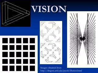
Topic 10 The Eye & the Sensory Modality of Vision Lange. Biology 463 - Neurobiology. Introduction. Significance of vision Relationship between human eye, camera, and projector screen Retina – the neural portion of the sense of vision

E N D
Topic 10 The Eye & the Sensory Modality of Vision Lange Biology 463 - Neurobiology
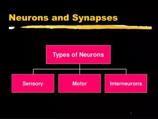
Introduction Significance of vision • Relationship between human eye, camera, and projector screen • Retina – the neural portion of the sense of vision • Photoreceptors: Converts light energy into neural activity • Detects differences in intensity of light • Detects differences in wavelengths of light • Lateral geniculate nucleus (LGN) • the primary processing center for visual information received from the retina of the eye • The LGN is found inside the thalamus
START: Retina – the neural portion of the sense of vision • Photoreceptors: Converts light energy into neural activity • Detects differences in intensity of light • Detects differences in wavelengths of light
END: Lateral geniculate nucleus (LGN) • the primary processing center for visual information received from the retina of the eye • The LGN is found inside the thalamus
Properties of Light • Light • Electromagnetic radiation • Wavelength • Amplitude
Light properties: • Energy is proportional to frequency (e.g., gamma radiation and cool colors - high energy ) (e.g., radio waves and hot colors - low energy )
Optics • Study of light rays and their interactions • Reflection • Bouncing of light rays off a surface • Absorption • Transfer of light energy to a particle or surface • Refraction • Bending of light rays from one medium to another All of these factors will influence how the retinal cells perceive light energy.
Near UV light that is absorbed in the lens changes proteins there causing them to become cloudy (cataracts). • High intensity blue light focused on the retina can also induce a photochemical reaction which causes the light receptors in the eye to lose sensitivity. This is known as photo bleaching and mostly effects blue color vision. • Long exposure to bright sunlight can cause photobleaching type of vision loss.
The Structure of the Eye Gross Anatomy of the Eye • Pupil: Opening where light enters the eye • Sclera: White of the eye • Iris: Gives color to eyes • Cornea: Glassy transparent external surface of the eye • Optic nerve: Bundle of axons from the retina
The Structure of the Eye Ophthalmoscopic appearance of the eye:
Punctum Caecum - the place in the visual field that corresponds to the lack of light-detecting photoreceptor cells on the optic disc of the retina where the optic nerve passes through it. Often called the “Blind Spot”. • Tests for detecting the “Blind Spot” in the visual field: • Positive Blind Spot Test (Easiest test to detect/locate the blind spot.) • The next slide will show a standardized postive blind spot test. • This test will likely NOT work as a projected image on the screen due to size. Instead, if you wish to try out this test, you will need to print out the slide (in the 4 or 6 slide per page format). • Close one eye. • Place the paper on a tabletop or wall, and stare at the “+” while approximately 25 cm away. • Move your head slowly towards the paper, and there will be a point where the “dot” will no longer appear. That is the location of your blind spot.
Tests for detecting the “Blind Spot” in the visual field (continued): • Negative Blind Spot Test (Shows how the eye compensates for the blind spot.) • The next slide will show a standardized negative blind spot test. • This test will likely NOT work as a projected image on the screen due to size. Instead, if you wish to try out this test, you will need to print out the slide (in the 4 or 6 slide per page format). • Close one eye. • Place the paper on a tabletop or wall, and stare at the two bars while approximately 25 cm away. • Move your head slowly towards the paper, and there will be a point where the missing segment of the line will disappear. This is the location of your blind spot, and shows how the brain compensates for this missing segment by attempting to “fill in” the missing information.
Strabismus – the collective name for misalignments of the eyes with each other Exotropia – the condition as seen in the image where the woman has one eye that points outward compared to the other Esotropia – strabismus where the misaligned eye points inward Strabismus that arises in childhood will result in the condition of amblyopia (lazy eye) and results in a loss of depth perception that often can be permanent if not treated early enough. Strabismus that arises in adulthood can be an indication of a brain tumor or stroke. It can also arise due to an accident.
In the 1960s, studies showed that children with untreated strabismus would often perform poorly in school. If severe enough, the mixed signals received by the brain would end up resulting in the visual signal of the straying eye to be fully ignored and the signals from the eye no longer being perceived in the brain. Studies during the last five years have also shown that children with strabisumus of the exotropia variety have a much higher rate of developing psychiatric disorders. To learn more: Mohney et. al., 2008. Mental Illness in Young Adults Who Had Strabismus as Children. Pediatrics. 122:1033- 1038.
Cataracts - a clouding that develops in the crystalline lens of the eye or in its envelope, varying in degree from slight to complete opacity Two primary forms: Senile Cataract – most typical, seen in the majority of cases. The lens will first yellow, and then will tend to shrink and grow more opaque (see image to the right) Morgagnian Cataract – a cataract where the lens will liquify and rupture as it grows cloudy (see below)
How the cornea and lens are used to focus images onto the retina.
Accommodation is the ability of the lens to flex by growing flatter (stretching out) or relaxing (becoming more bulbous) which further refines the ability of the eye to focus. Age related stiffening of the lens with a resultant loss in near vision is called presbyopia.
Forms of visual deficits based upon shape issues with the eye. The corrective lens(es) are chosen based upon what sorts of assistance to diffraction of light is (are) needed.
George Wald 1906 – 1997 received the Nobel Prize in 1967 for discoveries concerning the primary physiological and chemical visual processes in the eye
Image Formation by the Eye The Pupillary Light Reflex • Connections between retina and brain stem neurons that control muscle around pupil • Continuously adjusting to different ambient light levels • Consensual • Pupil similar to the aperture of a camera
Image Formation by the Eye • The Visual Field • Amount of space viewed by the retina when the eye is fixated straight ahead
Visual Acuity • Ability to distinguish two nearby points • Visual Angle: Distances across the retina described in degrees
Microscopic Anatomy of the Retina • Direct (vertical) pathway: • Ganglion cells • Bipolar cells • Photoreceptors
Microscopic Anatomy of the Retina • Retinal processing also influenced lateral connections: • Horizontal cells • Receive input from photoreceptors and project to other photoreceptors and bipolar cells • Amacrine cells • Receive input from bipolar cells and project to ganglion cells, bipolar cells, and other amacrine cells
Microscopic Anatomy of the Retina • The Laminar Organization • Inside-out • Light passes through ganglion and bipolar cells before reaching photoreceptors
Photoreceptor Structure • Converts electromagnetic radiation to neural signals • Three main regions • Outer segment containing a variety of PLB membrane disks that contain photopigments that absorb light • Inner segment (containing the cell body) • Synaptic terminal • Types of photoreceptors • Rods and cones
Regional Differences in Retinal Structure • Varies from fovea to retinal periphery • Peripheral retina • Higher ratio of rods to cones • Higher ratio of photoreceptors to ganglion cells • More sensitive to light Think through logically…. How would these different patterns of distribution of rods and cones be related to a) scotopic (night) vision, b) photopic (day) vision, and c) central vision vs. peripheral vision?
Regional Differences in Retinal Structure • Cross-section of fovea: Pit in retina where outer layers are pushed aside • Maximizes visual acuity • Central fovea: All cones (no rods) • 1:1 ratio with ganglion cells • Area of highest visual acuity
Macula Lutea - an oval-shaped highly pigmented, yellow spot near the center of the retina of the human eye • it has a diameter of around 5 mm • is often histologically defined as having two or more layers of ganglion cells • near its center is the fovea, a small pit that contains the largest concentration of cone cells in the eye and is responsible for central, high resolution vision. The macula also • contains the parafovea (the area surrounding the fovea and containing both rods and cones) and perifovea (the area that is only cones, sometimes called the fovea centralis). • Because the macula is yellow in color it absorbs excess blue and ultraviolet light that enter the eye, and acts as a natural sunblock
Phototransduction • Phototransduction in Rods • light energy interacts with photopigment to produce a change in membrane potential • analogous to activity at G-protein coupled neurotransmitter receptor • but causes a decrease in second messenger
Phototransduction in Rods • Dark current: Rod outer segments are depolarized in the dark because of steady influx of Na+ • Photoreceptors hyperpolarize in response to light
More About Phototransduction in Rods • light activated biochemical cascade in a photoreceptor • the consequence of this biochemical cascade is signal amplification
Opsins are proteins and the retinal-binding visual pigments found in the photoreceptor cells in the retinas of eyes. Rhodopsin, also known as visual purple, is a pigment of the retina that is responsible for both the formation of the photoreceptor cells and the first events in the perception of light. Conversion of Opsin into Rhodopsin: Exposed to light, the pigment retinal immediately photobleaches, and it takes about 30 minutes to regenerate fully in humans.
Mutations in the rhodopsin gene is a major contributor to various retinopathies such as: • Retinitis Pigmentosa - the mutated gene produces proteins which are difficult to remove. This can disrupt the intermediate filament network of opsin and impairs the ability of the cell to degrade these non-functioning proteins. This can lead to premature photoreceptor apoptosis. • X-linked Congenital Stationary Night Blindness - has two forms, complete, known as type-1 (CSNB1), and incomplete, known as type-2 (CSNB2). The difference between each form is a matter of severity of effect. In the complete form (CSNB1), there are no measurable rod cell responses to light, whereas this response is mildly measurable in the incomplete form. Make note of the pigmented regions where vision is lost.
Phototransduction • Dark and Light Adaptation • Dark adaptation—factors • Dilation of pupils • Regeneration of unbleached rhodopsin • Adjustment of functional circuitry 20–25 minutes All-cone daytime vision All-rod nighttime vision
Young-Helmholtz Trichromacy Theory of Color Vision - there are three receptors in the retina that are responsible for the perception of color. One receptor is sensitive to the color green, another to the color blue, and a third to the color red. These three colors can then be combined to form any visible color in the spectrum.
Phototransduction in Cones • Similar to rod phototransduction • Three different opsins • Red, green, blue • Color detection • Contributions of blue, green, and red cones to retinal signal • Spectral sensitivity
Color Blindness - a genetic disorder where individuals lose the ability to distinguish some or all variations in color vision
Deuteranopia & Deuteranomaly (forms of GREEN deficiency) Deuteranopia and deuteranomaly are the most common forms of color-blindness. People with these conditions have cones that are insensitive to medium wavelengths (greens), but the end result is similar to protonopia, with the exception that reds do not look as dark. Deuteranomaly is the less severe of the two conditions. Individuals with deuteranomaly cannot see reds and greens in the same way that people with full color vision can, they are able to distinguish between shades of reds and greens relatively accurately. full color vision green deficiencies
Protanopia & Protanomaly (forms of RED deficiency) Color receptors in the eyes of people with protanopia are not sensitive to long wavelengths (the reds). Reds look more like beiges and appear to be somewhat darker than they actually are. The greens tend to look similar to the reds. Protanomaly is milder than protanopia, but the end result is similar. Although many people with protanomaly can distinguish some reds and greens, they cannot do so as easily as someone with color-normal vision, and, as with protanopia, reds tend to look darker as well. red deficiencies full color vision
Tritanopia (a form of BLUE deficiency) Note… Tritanopia is much less common than the other categories mentioned above. Tritanopia is insensitivity to short wavelengths (the blues). Blues and greens can be confused, but yellows are also affected in that they can blend with blues or appear as lighter shades of red. full color vision blue deficiencies
Rod Monochromacy or Achromacy (NO color vision) This group constitutes an extremely small minority among people who are color-blind. All cones of the eye are non-functional, so the rods (receptors which can only differentiate between light and dark) are the only available source of visual information. Individuals with achromacy see no color at all. People with achromacy often have poor visual acuity and have an aversion to bright light. full color vision complete cone deficiency