Download
1 / 24
240 likes | 1.15k Views
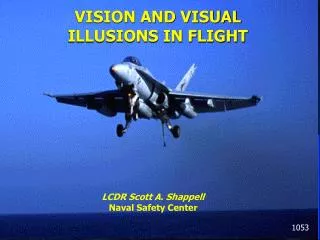
Special Senses: Vision. Chapter 8. Agenda. Anatomy of the eye – internal and external Light and light refraction Diseases and disorders of the eye. The Eye and Vision. Special sense receptors – large, complex sensory organ 70% of sensory receptors Optic nerve – 1+ million nerve fibers.

E N D
Special Senses: Vision Chapter 8
Agenda • Anatomy of the eye – internal and external • Light and light refraction • Diseases and disorders of the eye
The Eye and Vision • Special sense receptors – large, complex sensory organ • 70% of sensory receptors • Optic nerve – 1+ million nerve fibers
External and Accessory Structures of Eye • Extrinsic eye muscles Figure 8.3a
External and Accessory Structures of Eye • Eyelids – eyelashes • Tarsal glands - lubrication • Conjunctiva • Lacrimal Apparatus http://www.wisedude.com/health_medicine/eyelids.htm http://www.virtualmedicalcentre.com/anatomy.asp?sid=28
Conjunctivitis – Pink Eye • Conjunctivitis – inflammation of conjunctiva • Pink Eye – Infection of conjunctiva by bacteria or virus, highly contagious • Leads to red, irritated eyes https://www.bcbsri.com/BCBSRIWeb/images/mayo_popup/Pinkeye(conjunctivitis).jsp
Internal Anatomy of Eye Figure 8.4a • Humors • Vitreous - posterior • Aqueous – anterior • Lens • Wall – 3 layers • Fibrous layer • Vascular layer • Sensory layer Figure 8.4b
Wall of Eye • Fibrous Layer – 2 parts • Sclera – protective, white of eye • Cornea – crystal clear, many nerve endings, vulnerable, extraordinary healing • Only tissue in body that can be transplanted w/o fear of rejection – no blood vessels immune system can’t reach http://www.vision-training.com/en/Eye%20Anatomy/Eye%20anatomy.html
Wall of Eye • Vascular Layer – 3 regions • Choroid – blood rich, dark pigment, light scatter • Ciliary body – muscle structure, lens attached via ciliary zonule (suspensory ligament) • Iris – muscle structure, pigmented, rounded opening (pupil), smooth muscle http://canyouspellhubbell.com/wordpress/?p=15
Wall of Eye • Sensory Layer – 2 layer retina • Extends to ciliary body • Outer layer – Pigmented Layer • Pigmented cells – absorb and prevent light scattering • What other structure also does this? • Cells act as phagocytes Figure 8.5b
Wall of Eye • Sensory Layer – 2 layer retina • Extends to ciliary body • Inner Layer – Neural Layer • Transparent • Contains millions of photorectptors- cones and rods Figure 8.5
Nerve Impulses from Retina • Electrical signals from photoreceptors leave retina as nerve impulses via optic nerve • Transmitted to optic cortex = vision • Photoreceptors on entire retina except where optic nerve leaves eyeball = optic disk or blind spot
Photoreceptors • Cones – most dense at center of retina, decrease on periphery • Color under bright conditions • Macula lutea, fovea centralis • 3 types – sensitive to different wavelengths • Blue • Green • Green and Red (called red) • ‘mixing’ in brain http://pro.corbis.com/search/Enlargement.aspx?CID=isg&mediauid=%7B0CA81D50-A4C0-40AE-B9FF-351B3485A49E%7D
Photoreceptors • Rods – more dense at periphery, decrease in number towards center • See grey tones in dim light • Provides peripheral vision
Night or Color Blindness • Color Blindness – More common in men, most common is lack of red or green receptors • See red or green as same color, depends on cones present • Can see differences in intensity of color • Night Blindness – most commonly caused by Vitamin A deficiency, leads to deterioration of neural retina • Vitamin A supplements can restore if degenerative changes have not occurred
Lens • Divides eye into 2 segments • Anterior (Aqueous) Segment • Similar to blood plasma • Continuously secreted by part of choroid • Provides nutrients for lens and cornea, lack blood supply • Reabsorbed into venous blood through canal of Schlemm • Posterior (Vitreous) Segment • Gel-like substance • Both maintain intraocular pressure
Glaucoma • Drainage of aqueous fluid via canal of Schlemm blocked • pressure increases compressing retina and optic nerve, causes pain and possibly blindness • Common in elderly • Slow process http://www.steadyhealth.com/articles/Can_Glaucoma_Be_Prevented__a640_f0.html
Lens • Focuses light entering retina • Biconvex • Held upright by suspensory ligaments – ciliary zonule attached to ciliary body http://projects.cbe.ab.ca/Diefenbaker/Biology/Bio%20Website%20Final/notes/nervous_system/8_eye_notes.html
Light Refraction • Refraction = bending of light due to change in speed • Refractive power of cornea, and humors constant • Refractive power of lens changes • Goal to focus light at one point on back of eye • How does lens change to do this? • Distant vision – light enters as parallel rays, lens does not need to change shape • Close vision – divergent rays, lens must bulge more = accommodation
Cataracts • With age, lens becomes hard and opaque • Diabetes mellitus • Heavy smoking • Vision becomes hazy and distorted, will lead to blindness • Surgical removal or glasses http://www.ehponline.org/docs/2005/113-3/niehsnews.html
Binocular vision • “two-eyed” vision • Provides depth perception • 3D vision http://www.e-advisor.us/visual_fields.htm
Eye Reflexes • Convergence = movement of eyes medially when viewing close objects • Photopupillary reflex = pupils constrict in bright light to protect photoreceptors • Accommodation pupillary reflex = pupils constrict when viewing close objects