Download
1 / 27
280 likes | 391 Views
Nerve Signal Transmission. Raise your right hand. Easy, right? You don’t even have to think twice and your right arm is moving…. But what makes it happen??? How does your brain tell your body what to do?. The Nervous System. Central nervous system (CNS) brain and spinal cord

E N D
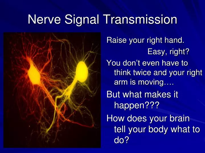
Nerve Signal Transmission Raise your right hand. Easy, right? You don’t even have to think twice and your right arm is moving…. But what makes it happen??? How does your brain tell your body what to do?
The Nervous System • Central nervous system (CNS) • brain and spinal cord • Peripheral nervous system (PNS) • sensory and motor neurons • Nerves • bundles of neurons wrapped in connective tissue
Simple Nerve Circuit • Reflex: simple response-- sensory to motor neurons • Involuntary; not analyzed or interpreted by brain • Ganglion (ganglia): cluster of nerve cell bodies in the PNS • Glia: cell that provides support, insulation, and protection • astrocytes, radial glia, oligodendrocytes, Schwann cells
Information Processing • External stimuli detected • sight, sound, touch, smell, taste, etc. • Information sent to the CNS • analysis and interpretation • Motor output is relayed to the effector cells • muscle/ endocrine cells
Neuron Structure • Cell body~ nucelus and organelles • Dendrites~ receiving signals • Axons~ transmitting signals • Hillock~ connects axon to cell body; generates signal • Synaptic terminals~ communicates w/ another cell (releases neurotransmitters) • Synapse~ neuron junction (site of cell communication)
Neuron Diversity • Sensory neuron: convey information to spinal cord • Interneurons: information integration • Motor neurons: convey signals to effector cell (muscle or gland)
Schwann Cells and Myelin • Schwann cells • PNS support cells • Wraps around axon, creating layers of myelin • Myelin sheath • supporting, insulating layers • Nodes of Ranvier • Gaps between Schwann cells
Membrane Potential • Intracellular/extracellular ionic concentration difference • K+ diffuses out/Na+ in; large anions cannot follow (selective permeability of the plasma membrane) • Voltage difference between -60 and -80 mV
It has potential…. • Resting potential~ membrane potential of a neuron that is not transmitting • Equilibrium potential~ magnitude of membrane voltage at equilibrium • Nernst equation- Eion = 62mV (log [ion]outside / [ion]inside
Gated Ion Channels Open/ Close in response to stimuli… • Photoreceptors • Changes in light intensity • Vibrations in air • sound receptors • Chemical • neurotransmitters • Voltage • membrane potential changes
Graded Potentials • Depend on strength of stimulus • Threshold potential must be reached for reaction to occur • Hyperpolarization (outflow of K+); increase in electrical gradient; cell becomes more negative • Depolarization (inflow of Na+); reduction in electrical gradient; cell becomes less negative
Threshold potential: if stimulus reaches a certain voltage (-50 to -55 mV)….The action potential is triggered…. 1. Resting state • Both Na+ and K+ voltage-gated channels are closed 2. Threshold • a stimulus opens some Na+ channels 3. Depolarization • action potential generated • Na+ channels open • cell becomes positive (K+ channels closed) 4. Repolarization • Na+ channels close, K+ channels open; K+ leaves • cell becomes negative 5. Undershoot • both gates close, but K+ channel is slow; resting state restored • Refractory period~ insensitive to depolarization due to closing of Na+ gates
Conduction of Action Potential • Movement of the action potential is self-propagating • Depolarization of one part of axon triggers the action potential in the next part • Regeneration of “new” action potentials only after refractory period • This keeps signal moving in the forward direction only
Action Potential Speed • Axon diameter (larger = faster; 100m/sec) • Nodes of Ranvier (concentration of ion channels) • saltatory conduction- transmission “jumps” from one node to the next • 150m/sec
Synaptic communication • Depolarization of the membrane (from a.p.) causes Ca+ influx (voltage gated channel) • Ca+ causes vesicles to fuse with the presynaptic membrane and release neurotransmitters • Neurotransmitters bind w/ ligand-gated ion channels on postsynaptic membrane • Neurotransmitter releases from the receptor and the channels close
Postsynaptic Potential • EPSPs- excitatory postsynaptic potentials • IPSPs- inhibitory postsynaptic potentials
Indirect Transmission • Neurotransmitters bind with specific receptors instead of ion channels • Much slower reaction time but last longer • Different neurotransmitters produce diverse effects • Psychological drugs interact at the location of these receptors
Muscles contract • Muscles move by shortening, or contracting… they cannot extend on their own • Each muscle has an antagonistic muscle that contracts to move in the opposite direction
What’s in a Muscle? • Muscles are a bundle of muscle fibers. • Muscle fibers: single cell w/ many nuclei composed of myofibrils • Myofibrils: longitudinal bundles composed of myofilaments • Myofilaments: • Thin~ 2 strands of actin protein and a regulatory protein • Thick~ myosin protein • Sarcomere: repeating unit of muscle tissue within a myofibril
Sarcomere Structure • Z lines~ sarcomere border • I band~ only actin • A band~ actin and myosin overlap • H zone~ central sarcomere; only myosin
Sliding-filament model • Theory of muscle contraction • Sarcomere length reduced • Z line length becomes shorter • Actin and myosin slide past each other (overlap increases)
Actin-myosin interaction • Myosin head hydrolyzes ATP to ADP and inorganic phosphate (Pi)-- “high energy configuration” • Myosin head binds to actin; forming a “cross bridge” • Releasing ADP and P, myosin relaxes sliding actin; “low energy configuration” • Binding of new ATP releases myosin head • Creatine phosphate~ supplier of phosphate to ADP
Contraction Regulation • Relaxation: • tropomyosin blocks myosin binding sites on actin • Contraction: • calcium binds to toponin complex • tropomyosin changes shape, exposing myosin binding sites
ACTION! • Acetylcholine released from synaptic terminal of motor neuron • Acetylcholine binds w/ receptors and causes an action potential in the muscle fiber • A.P travels down the T (transverse) tubules and triggers release of Ca+ from the sarcoplasmic reticulum • Modified endoplasmic reticulum • Contraction begins when Ca+ reaches the sarcomere and binds to troponin