Download
1 / 46
460 likes | 571 Views
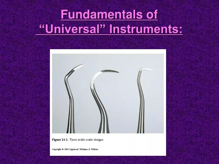
Fundamentals of “Universal” Instruments:. To date you’ve learned: *#17 Explorer. To date you’ve learned: -Gracey 1/2 -204s. 2 Types of Universal Instruments:. 1. Scalers - (in cross-section) pointed tip pointed back supragingival calculus removal

E N D
To date you’ve learned: *#17 Explorer To date you’ve learned: -Gracey 1/2 -204s
2 Types of Universal Instruments: 1. Scalers - (in cross-section) pointed tip pointed back supragingival calculus removal 2. Curets - (in cross-section) rounded tip rounded back sub/supragingival calculus removal
Types of Sickle Scalers: • Anterior- ‘straight’ shank instrument • Posterior- ‘curved’ or multiple shank instrument • (actually can be used universally in the mouth)
Top instrument (Unpaired) is an example of a curet (on the left) and an anterior sickle on the (right) Bottom instrument (paired) is an example of a posterior sickle on both ends.
Design Characteristics: Basically there are 2 cutting edges at each end. The Face of the Sickle is at a 90 degree angle to the terminal shank. Flat “face” Terminal shank
Sickle instrument have two cutting edges on each end of the instrument:
Terminal shank is at a 90 degree angle to the Face
Face Face
Your instrument casetes do not include the Anterior Sickle. • You will use the Posterior Sickle throughout the mouth including the anterior teeth.
P. 210 p.290 In Nield Book (mirror, sickle, etc.) (mirror) (sickle)
Keep In Mind: • Operator and Client Positioning • Instrument Blade Selection • Grasp • Fulcrum • Insertion • Adaptation • Angulation
Please remember: The Sickle instrument is used SUPRAGINGIVAL! (You can go sub about 1-2 mm if necessary, but not more than that!!!)
Anterior Scaler Initial point of insertion is always at the line angle The above shows a straight shanked sickle. Since we will not use this type of instrument, please focus instead on the Terminal Shank and its cutting edges and how it relates to the tooth.
Mandibular Anteriors: BuccalRight Handed Clinician 5 6 4 3 1 2 #27 #26 #25 #24 #23 #22 • From a 12:00 position, insert at the Distal Buccal Line Angle of #22 (tip towards the col) • Initiate walking stroke towards and into the distal col. Remove at end of pull stroke • Reinsert at the Mesial Buccal Line Angle of #22 (tip towards the mesial col) • Initiate walking stroke toward and into the mesial col. Remove at end of pull stroke • Move onto #23 D and then #23 M, then #24D, #24 M, etc.
Mandibular Anteriors: Lingual Right Handed Clinician #22 #23 #24 #25 #26 #27 • From a 12:00 position, insert at the Distal Lingual Line Angle of #22 (tip towards the col) • Initiate walking stroke towards and into the distal col. Remove at end of pull stroke • Reinsert at the Mesial Lingual Line Angle of #22 (tip towards the mesial col) • Initiate walking stroke toward and into the mesial col. Remove at end of pull stroke • Move onto #23 D and then #23 M, then #24D, #24 M, etc.
Mandibular Anteriors: BuccalLeft Handed Clinician 5 6 4 3 1 2 #27 #26 #25 #24 #23 #22 • From a 12:00 position, insert at the Distal Buccal Line Angle of #27 (tip towards the col) • Initiate walking stroke towards and into the distal col. Remove instrument at upward stroke • Reinsert at the Mesial Buccal Line Angle of #27 (tip towards the mesial col) • Initiate walking stroke toward and into the mesial col. Remove instrument at upward stroke • Move onto #26 D and then #26 M, then #25D, #25 M, etc.
Mandibular Anteriors: Lingual Left Handed Clinician #22 #23 #24 #25 #26 #27 • From a 12:00 position, insert at the Distal Lingual Line Angle of #27 (tip towards the col) • Initiate walking stroke towards and into the distal col. Remove at end of pull stroke • Reinsert at the Mesial Lingual Line Angle of #27 (tip towards the mesial col) • Initiate walking stroke toward and into the mesial col. Remove at end of pull stroke • Move onto #26 D and then #26 M, then #25D, #25 M, etc.
1 2 1 2 1 2 1 2 Notice that you will begin your working stroke at the Distal Line Angle(1) and proceed into the distal col. You will then reinsert tip at the Mesial Line Angle(2) (tip now pointing towards the Mesial). Remember, Terminal shank of instrument is parallel to the line angle- proceed with walking stroke into the mesial col area.
Correct: Auxiliary Shank Terminal Shank
Incorrect: Auxiliary Shank Terminal Shank
Do Notuse the sickle on direct buccal/lingual surfaces!!!
Walking Sequence (per quadrant): (limited radius) Midline 2. 1. 2. 2. 2. 2. 1. 2. 2. For Right Handed Clinicians: 2. 2. 2. 1. 2. 2. Notice that operatorposition and the direction of the instrument handle changes at the canine on the dominant side. 1. 2. 2. 1. 1. 2. 1. 2. 1. 2. 1. 2. 2. 1.
Walking Sequence (per quadrant): (limited radius) Midline 1. 2. 2. 2. 1. 2. 2. For Left Handed Clinicians: 2. 1. 2. 2. 2. 1. 2. 2. Notice that operatorposition and the direction of the instrument handle changes at the canine on the dominant side. 1. 2. 1. 2. 1. 2. 1. 2. 1. 2. 1. 2. 1. 2.
So, what are we saying? You will be using the Posterior Sickle on ALL proximal surfaces in the mouth. When you are scaling only the anterior teeth- please follow the sequence in slides (slides #20-23) When scaling posterior and anterior teethin a single session- instrumentation sequence is: quadrant at a time! (slides #35/36)
In Review You Should ALWAYS Keep in Mind: • Lateral Pressure • Strokes • Stroke Direction • Stroke Length • Reinforcement
Now, Let’s take a look at the Universal Curet
The Universal curet is similar to the Sickle in that it also has two cutting edges per end. • The difference between them is that the tip of the Universal curet is rounded- not pointed like the Sickle. • This allows you to use this instrument SUBGINGIVAL!!!
Universal Curets: Scalers - (in cross-section) • pointed tip pointed back supragingival calculus removal Curets - (in cross-section) rounded tip rounded back sub & supragingival calculus removal
Universal Curets • Columbia • Barnhardt • Younger Good • Blade size, shank length and design will determine preferred area usage.
So, Let’s Keep in Mind. . . • Fulcrum rest must be near, but not directly over the surface being scaled (fulcrum on same arch). • Determine correct working end of instrument: • Terminal shank parallel to MESIAL line angle of molar • Foot of instrument curves towards the tooth • Angle for insertion is 0-40 (closed blade)
Continued. . . . • With closed blade, insert subgingival to JE- lateral pressure should be fairly light. • Open angle to 45-90° (subgingival) and initiate exploratory stroke (pull stroke) • Lateral pressure against tooth should remain fairly light unless a ‘bump’ is felt. Return back to JE and apply firmer pressure during pull stroke to remove ‘bump’ calculus? • Fulcrum pressure increases during pull stroke
Continued. . . • Relax fingers during exploratory stroke • Apply greater lateral pressure during pull stroke. • Strokes should be short and controlled, with moderate pressure from the base of the pocket, toward the gingival margin. • Stroke direction – vertical, oblique & horizontal (overlapping) remaining primarily in the gingival sulcus.
Please remember~ • You should ALWAYS scale a tooth to completion!!! • That means: Look at the clock and budget your time!!! • Quesstimate how long it will take you to scale a lingual/buccal surface • Dependant on : How ‘heavy’ the calculus is • How ‘tenacious’ the calculus is • How good is your technique?!? • Then determine how many teeth you can scale thoroughly and completely with the amount of time you have. . .