Download
1 / 39
1.46k likes | 4.88k Views
Front of thigh. Surface landmarks- Iliac crest Anterior superior spine Inguinal ligament Pubic tubercle Pubic symphysis Greater trochanter Patella Tibial tuberosity Ligamentum patella Medial & lateral condyles of femur and tiba Adductor tubercle. Front of thigh. Superficail fascia-

E N D
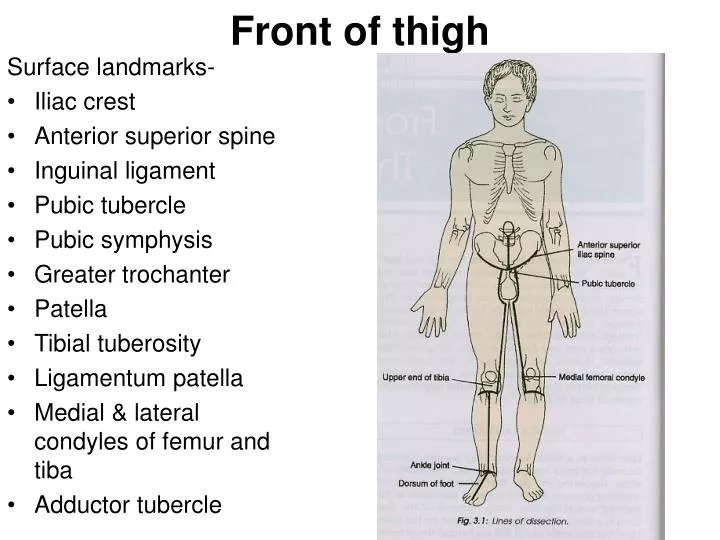
Front of thigh Surface landmarks- • Iliac crest • Anterior superior spine • Inguinal ligament • Pubic tubercle • Pubic symphysis • Greater trochanter • Patella • Tibial tuberosity • Ligamentum patella • Medial & lateral condyles of femur and tiba • Adductor tubercle
Front of thigh Superficail fascia- • Superficial fatty layer • Deep membranous layer • Holden’s line
Front of thigh Cutaneous nerves- 7 cutaneous nerves Lateral cutaneous branch of the subcostal nerve • Ilioingunial nerve(L1) • Femoral branch of the genitofemoral nerve(L1,2) • Lateral cutaneous nerve of thigh(L2,3) • Intermediate cutaneous nerve of thigh(L2,3) • Medial cutaneous nerve of thigh(L2,3) • Saphenous nerve(L3,4)
Front of thigh Patellar plexus • Ant div of the lateral cut nerve • Intermediate cut nerve of thigh • Ant div of the medial cut nerve
Front of thigh Cutaneous arteries- • Superficial external pudendal a • Superficial epigastric a • Superficial circumflex iliac a
Front of thigh Great saphenous vein-
Front of thigh Superficial inguinal lymph nodes-
Front of thigh Subcutaneous bursa- • Prepatellar bursa • Subcutaneous infrapatellar bursa
Front of thigh Deep fascia(fascia lata)-
Front of thigh Iliotibial tact- Insertion of gluteus maximus & tensor fascia latae
Front of thigh Saphenous opening-
Femoral triangle Boundaries- Laterally- medial border of sartorius Medially- medial border of adductor longus Base- inguinal ligament Apex-convergence of medial & lateral border
Femoral triangle Roof- • Skin • Superficial fascia containing superficial inguinal lymph nodes • Femoral branch of genitofemoral nerve • Branches of ilioinguinal nerve • Superficial branches of femoral vessels • Upper part of great saphenous vein • Deep fascia with saphenous opening & cribriform fascia
Femoral triangle Floor- Medially-adductor longus & pectineus Laterally-psoas major & iliacus
Femoral triangle Content • Femoral artery & its branches • Femoral vein & its tributaries • Great saphenous vein • Femoral sheath • Femoral nerve • Nerve to pectineus • The femoral branch of the gentiofemoral nerve • Lateral cut nerve of thigh • Deep inguinal lymph nodes
Femoral triangle Femoral sheath Boundaries- Divided into 3 compartments • Lateral or arterial compartment( femoral a & the femoral branch of genitofemoral nerve) • Intermediate or venous compartment( femoral vein) • Medial or lymphatic compartment( femoral canal)
Femoral triangle Femoral cana(1.5cm l &1.5 cm base)- Boundaries of femoral ring- • Ant-inguinal lig • Post- pectineus & its covering fascia • Med- concave margin of lacunar ligament • Lat-septum separating it from femoral vein • Femoral septum • Femoral fossa Content- lymph node of Cloquet or Rosenmuller & small amount of aerolar tissue
Femoral triangle Femoral hernia- • Typical course of hernial sac • Strangulation hernia
Femoral triangle Femoral artery- Origin- Course & extent Branches- • Superficial epigastric • Superficial external pudendal • Superficial circumflex iliac • Deep external pudendal • Profunda femoris • Muscular branches
Femoral triangle Relations-
Femoral triangle Profunda femoris artery- Course & relations Branches- • Medial circumflex femoral a • Lateral circumflex femoral a
Femoral triangle Femoral vein
Femoral triangle Femoral nerve- origin & root value- Course Branches- Muscular- ant supplies- sartorius & post supplies all the vasti and rectus femoris Cutaneous- ant div gives 2 cut ie intermediate & medial cut nerve of thigh and post div gives 1 ie saphenous nerve
Femoral triangle Articular branches- hip jt & knee jt Vascular branches to femoral artery & its branches
Adductor canal Extent- Shape- Boundaries- Ant- vastus medialis Post ( floor)- adductor longus above & adductor magnus below Med-strong fibrous membrane joining ant & post wall & over lapped by sartorius
Adductor canal Subsartorial plexus- • Medial cut nerve of thigh • Ant div of obturator nerve • Saphenous nerve
Adductor canal Contents- • Femoral artery • Femoral vein • Saphenous nerve • Nerve to vastus medialis • Two div of obturator nerve ie ant & post div
Muscles of anterior compartment of thigh Sartorious- Origin-ant sup iliac spine & upper half of the notch below the spine Insertion- upper part of the medial surface of the shaft of the tibia in front of the insertion of the gracillis & semitendinosus Nerve supply-femoral nerve Action-abductor & lateral rotator of the thigh and flexor of leg. ( palthi)
Muscles of anterior compartment of thigh Quadriceps femoris- Rectus femoris Origin-straight head from upper half of the ant inf iliac spine & reflected head from the groove above the margin of the acetabulum and capsule of the hip jt Insertion- base of the patella Nerve supply- femoral nerve Action- flexes the hip jt along with iliopsoas and helps to maintain the erect posture
Muscles of anterior compartment of thigh Vastus lateralis- Origin- upper part of intertrochanteric line, ant & inf borders of the greater trochanter, lateral lip of gluteal tuberosity & upper half of the lateral lip of linea aspera Insertion- lat part of the base of patella, upper 1/3rd of the lat border of patella & to the capsule of knee jt & iliotibial tract Nerve supply- femoral nerve Action- extensor of the knee jt
Muscles of anterior compartment of thigh Vastus medialis- Origin-lower part of intertrochanteric line, spiral line, medial lip of linea aspera & upper 1/4th of the medial supracondylar ridge Insertion-medial 1/3rd of the base of the patella & upper 2/3rd of the medial border of the patella Nerve supply- femoral nerve Action-extensor of knee jt and prevents lateral displacement of the patella
Muscles of anterior compartment of thigh Vastus intermedius- Origin- upper 3/4th of ant & lat surfaces of the shaft of the femur Insertion-base of the patella Nerve supply- femoral nerve Action-extensor of the knee jt