Download
1 / 45
550 likes | 951 Views
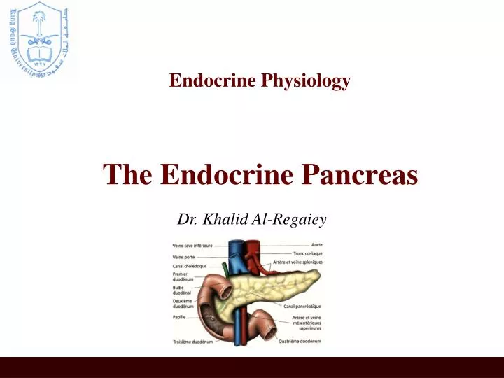
Endocrine Physiology The Endocrine Pancreas. Dr. Khalid Al-Regaiey. Pancreas. A triangular gland, which has both exocrine and endocrine cells, located behind the stomach Strategic location Acinar cells produce an enzyme-rich juice used for digestion (exocrine product)

E N D
Endocrine PhysiologyThe Endocrine Pancreas Dr. Khalid Al-Regaiey
Pancreas • A triangular gland, which has both exocrine and endocrine cells, located behind the stomach • Strategic location • Acinar cells produce an enzyme-rich juice used for digestion (exocrine product) • Pancreatic islets (islets of Langerhans) produce hormones involved in regulating fuel storage and use.
Islets of Langerhans • 1-2 million islets • Beta (β) cells produce insulin (70%) • Alpha (α) cells produce glucagon (20%) • Delta (δ) cells produce somatostatin (5%) • F cells produce pancreatic polypeptide (5%)
Insulin • Hormone of nutrient abundance • A protein hormone consisting of two amino acid chains linked by disulfide bonds • Synthesized as part of proinsulin (86 AA) and then excised by enzymes, releasing functional insulin (51 AA) and C peptide (29 AA). • Has a plasma half-life of 6 minutes.
Insulin Synthesis DNA (chromosome 11) in β cells mRNA Preproinsulin (signal peptide, A chain, B chain, and peptide C) proinsulin insulin
Insulin Synthesis • Insulin synthesis is stimulated by glucose or feeding and decreased by fasting • Threshold of glucose-stimulated insulin secretion is 100 mg/dl. • Glucose rapidly increase the translation of the insulin mRNA and slowly increases transcription of the insulin gene
Blood glucose concentration Gastrointestinal hormones Blood amino acid conc. Major control Food intake Islet cells Sympathetic stimulation (and epinephrine) Parasympathetic stimulation Insulin secretion Factors controlling insulin secretion Blood glucose Blood fatty acids Blood amino acid Protein synthesis Fuel storage
Insulin Receptor • the insulin receptor is a transmembrane receptor • belongs to the large class of tyrosine kinase receptors • Made of two alpha subunits and two beta subunits
Raapid (seconds) • (+) transport of glucose, amino acids, K+ into insulin-sensitive cells • Intermediate (minutes) • (+) protein synthesis • (-) protein degradation • (+) of glycolytic enzymes and glycogen synthase • (-) phosphorylase and gluconeogenic enzymes • Delayed (hours) • (+) mRNAs for lipogenic and other enzymes
Action of insulin on Adipose tissue • (+) glucose entry • (+) fatty acid synthesis • (+) glycerol phosohate synthesis • (+) triglyceride dep0sition • (+)lipoprotein lipase • (-) of hormone-sensitive lipase • (+) K uptake
Action of insulin on Muscle: • (+) glucose entry • (+) glycogen synthesis • (+) amino acid uptake • (+) protein synthesis in ribosomes • (-) protein catabolism • (-) release of gluconeogenic aminco acids • (+) ketone uptake • (+) K uptake
Action of insulin on Liver: • (-) ketogenesis • (+) protein synthesis • (+) lipid synthesis • (-)gluconogenesis, (+) glycogen synthesis, (+) glycolysis.
General • (+) cell growth
Glucose Transport • GLUT1 (erythrocytes, brain) • GLUT2 (liver, pancreas, small intestines) • GLUT3 (brain) • GLUT4, insulin sensitive transporter (muscle, adipose tissue)
Glucagon • A 29-amino-acid polypeptide hormone that is a potent hyperglycemic agent • Produced by α cells in the pancreas
SYNTHESIS DNA in α cells (chromosome 2) mRNA Preproglucagon proglucagon glucagon
Glucagon Actions • Its major target is liver: • Glycogenolysis • Gluconeogenesis • Lipid oxidation (fully to CO2 or partially to produce keto acids “ketone bodies”). • Release of glucose to the blood from liver cells
Diabetes • Diabetes is probably the most important metabolic disease. • It affects every cell in the body and affects carbohydrate, lipid, and protein metabolism. • characterized by the polytriad: • Polyuria (excessive urination) • Polydypsia (excessive thirst) • Polyphagia (excessive hunger).
Types of Diabetes Type 2diabetes Affects adults Causedefect in insulin action Treatment : diet or OHA Type 1Diabetes Affects children Cause:inadequate insulin secretion Treatment :insulin injection
Diabetes Mellitus Cause Inadequate secretion of insulin Defects in the action of insulin Metabolic disturbances (hyperglycemia and glycosuria)
Diabetes Mellitus Type I • Caused by an immune-mediated selective destruction of β cells • β cells are destroyed while α cells are preserved: No insulin :::: high glucagon high production of glucose and ketones by liver glucose & ketones osmotic diuresis keto acids diabetic ketoacidosis
Diabetes Mellitus: Type II • More common in some ethnic groups • Insulin resistance keeps blood glucose too high • Chronic complications: atherosclerosis, renal failure & blindness
Glucose Tolerance Test • Both the FPG and OGTT tests require that the patient fast for at least 8 hours (ideally 12 hr) prior to the test. • The oral glucose tolerance test (OGTT): • FPG test • Blood is then taken 2 hours after drinking a special glucose solution
Glucose Tolerance Test (GTT) • Following the oral administration of a standard dose of glucose, the plasma glucose concentration normally rises but returns to the fasting level within 2 hours. • If insulin activity is reduced, the plasma glucose concentration takes longer than 2 hours to return to normal and often rises above 200 mg/dl. • Measurement of urine glucose allows determination of the renal threshold for glucose.
Glucose Tolerance Test • The following results suggest different conditions: • Normal values: • FPG <100mg/dl • 2hr PPG < 140 mg/dL • Impaired glucose tolerance • 2hr PPG = 140 - 199 mg/dL • Diabetes • FPG ≥ 126 mg/dl • 2hr PPG levels ≥200 mg/dL