Download
1 / 33
330 likes | 518 Views
Hypersensitivity. Lecture 12. Hypersensitivity – allergy immunedeficiency. Deficiency of regulatory function immune system Occasionally even healthy immune system react not correctly – hyperactively 1963 – Coombs a Gell classifiction

E N D
Hypersensitivity Lecture 12
Hypersensitivity – allergy immunedeficiency Deficiency of regulatory function immune system Occasionally even healthy immune system react not correctly – hyperactively 1963 – Coombs a Gell classifiction acc.to the type of ethiological immune mechanism Worldwide , influenced Sensitivity is – individual (genetic,exposition – smoking, home pets) immunological base of some clinical sy is not always clearly defined – intolerance, opium, intoxication by sea fruits - dif.dg
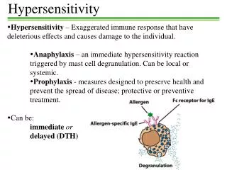
Terms • Allergy – disease, during which the immune reaction to the antigen (alergen) is based on inflamation of tissues that leads to the changes of the function without elimination of antigen • Allergen – antigen starting allergic reaction – chemicals – natural or synthetic • Different types of chemicals – different mechanism of allergy • HyperSenzitivita – too strong answer to antigen • Exposition to allergen • Types and phases of allergic reactions • Atopic – person prepared to react in allergic – hypersensitive way via IgE
Types of alergic-hypersensitive reactions Acc. Coombs and Gell – based on immunological mechanisms 4 types: 1. type – early reaction, atopy 2. type – cytotoxic reaction 3. type – reaction from immunocomplexes 4. type – delayed type hypersensitivity reaction - DTH • Possibility of existence of several reactions together
Phases of hypersensitive reactions A) Sensibilisation: Exposition to antigen induce stage (with specific antibodies or sensibilised lymphocytes), that is clinically seen only after the next exposition to the same antigen – the time of sensibilisation is not always known B) Fase of activity: • reexposition, • binding of specific effectors (Ab, cells) • production and release of cytokines, • clinical symptomatology
Immunological way 1) IgE-mastocyt-mediators - early reaction (type 1) 2) IgG or IgM-complement-neutrofil - cytotoxic reaction (type 2) - reaction from immunokomplexes (type 3) 3) Sensibilised effektor Tlymohocyte - TDH delayed hypersensitivity – lymfokines - delayed type, cell reaction (type 4)
SENSIBILISATION Ag and APC Processing of antigen Contact with CD4 TH cells TH1?TH2 B cells change Production of to plasma cell sensiibilised TDH cells Productionof IK B) EFFECTOR PHASE 1 2,3 4 Alergen + + IgE + IgG/IgM + Tbb Mastocyt complement mediatores anafylatoxines lymfokines C5 clinical symptoms
Structure of antibodies 2 identical heavy and 2 identical light chains disulfidic bound - between heavy and light – between 2 heavy - inter-chain - intra-chain Variable and constant regions existence of hypervariable regions- HVR (or complementarity determining regions.Ab with different specificity have different HVR regions – regions with variaty of amino acids, that represent direct contact with Ag flexible joint region - Domains – nor heavy nor light are flat.,
IgE • Monomer • Exceptional structure - similar to IgG – monomer bit on H chain in constant region there are 3 domains (Fcepsilon 2-4) Fcepsilon 2 and 3 bind on specific receptors of mastocytes receptor mastocytov (FcepsilonRI)
1.type – early reaction – atopic (4 fázy) Sensibilisation Exposition to antigen results in antibody answer – synthesis of specific IgE antibodies Why and when IgE and atopy? Way of enter – the same Type of molecule – the same The size of the dose is important – very small amout of Ag General characteristic of immunity – past contacts, stimulation of immunity, exposition to ag in postnatal period (type Th1 or Th2, theory of hygiene)
1.type – early reaction (4 fázy) cont. • IgE adhere by their Fc fragment on the surface of mastocytes, that are in circulation • Second exposition to ag (10-14 days later) – - immediate bound to Fab fragment of IgE present on mastocytes, - degranulation of vasoactive amines (histamín, sérotonín), lipid mediators, chemotactic substances • Clinical symptomatology caused by effect of mediators (smooth musles contraction, vasodilatation, increased vessel permeability, bronchospasm, oedema, anaphylactic shock) • Allergy to drugs, molds, insect bite • Tested by – i.c. application
Contact of the mucous membrane – phagocyosis by macrophages – presentation to Thelper cells – IL2- and clonal selection – influence and help B cells to change to plasma cells producing IgE – Thelper cells are controled by Tsupresor cells. In atopics the deficiency of controlled mechanism – lack of supressors lead to hyperproduction of IgE
IgE are bound on basofils in tissue and mastocytes After reexposition – the alergen is bound to Fab fragment of IgE bound on the mastocyte, that leads to the production of signal and degranulatio of vasoactive amines from the mastocyte, that has the clinical effect (increase of permeability, brnchospasm...).
2.type – cytotoxic reaction • Antibodies against antigens on the surface of self cells or tissues as the result of - deficiency of tolerance • cross reactivity of antibodies against exogenous antigens, that have similar structure as self cell superficial structures • bound of foreign antigens on the surface of self cells • Mechanism of destruction: opsonisation of antigen on the surface of the cell with IgG, NK cells are bound by Fc fragment of antibody and cause lysis of cell, activation of complement and lysis of the cell • Examples: Pemfigus – IgG antibodies react with intracellular substances between epidermal cells Haemolytical anaemia – ex.: PNC, Goodpastureov sy , Morbus hemolyticus neonatorum, posttransfusion reaction, cold aglutinins, early rejection of graft
Drug induced haemolytical anaemia: Antigen (PNC) is bound on erytrocytes. Antibodies IgG/IgM are specifically bound that activates C1q leading to lysis of the cell - haemolysis
3.type hypersensitivity from immunocomplexes • Circulating antibodies produce in time of sensibilisation react with free antigen (present in blood in time of reexposition) that leads to the production of circulating immunocoza vzniku cirkulujúcich imunokomplexov (CIK) • They are usually eliminated by macrophages • The disease strarts if CIK are deposited in tissue and/or if complement is activated (production of anaphyltoxins and chemoattractants) • Usually after chronical exposition ot antigen – longlasting load of macrophages(chronical streptococcal infection, autoimmune diseases, LE, alergic alveolitis) • Depends on the type of Ag an Ab and size of immunocomplexes (small are in circulation, big are eliminated by macrophages, medium can be deposited)
Arthus reaction – local reaction of immunocomplexes – test for existence or risk of the 3rd type of hypersensitivity (serzm serum disease) Injected antigen + IgG = IK –– activation of complement, agregation of trombocytes, release of mediators, increase of permeability of vessels, local oedem, PMNL Value of CIK – activity of the disease (40j normal value)
4. type - delayed type hypersensitivity • 1.- 3. type are expressed within minutes after reexposition (20 minutes – to several hours) • 4. type – after more that 12 hrs – late type – there are 4 clinical subtypes a) Jones Mote b) kontact allergy c) tuberculin reaction d) granulomatouse reaction Prejavia sa za 1 – 3 dni za 14 a viac dní
Jones Mote – cutaneous basofil hypersensitivity • Very inferquent • Locel reaction • Basofilic infiltration under epidermis • Induced by soluble antigens • After 24 hrs oedem of the skin that persists one week
Kontaktná alergia, dermatitída, kontaktný ekzém • Po kontakte kože s alergénom • MX po 2-3 dňoch • Nízkomolekulárne haptény –prenikajú kožou – viažu sa na telesné proteiny –senzibilizácia, –aktivita Langerhansových buniek: fungujú ako fagocyty, tkanivové makrofágy APC – uvoľnenie lymfokínov, mononukleárny infiltrát, pľuzgieriky epidermálna reakcia
Tuberkulínová reakcia • Robert Koch Subkutánna aplikácia tuberkulínu (lipoprotein mykobaktérií) pocit ochorenia s teplotou a lokálny edém a indurácia Dnes: presne odmerané množstvo na testovanie špecifickej imunity proti mykobaktériám Do 24 hodinách edém a indurácia 15 mm infiltrácia monocytmi a makrogágmi. Pri pretrvávaní antigénnej expozície – až granulomatózny zápal Anergia – negatívna tuberkulínová reakcia napriek expozícii (pri aplikácii spolu s kortisonom)
Mantoux test • 0 – 15 mm – anergia (neprítomnosť špecifickej bunkovej imunity) preočkovanie • 15 mm – primeraná reakcia, edém a indurácia • 15 a viac – hyperergia – pravdepodobná kontinuálna stimulácia - existencia expozície antigénu – ochorenie, kontakt – depistáž, rtg aj v okolí.
Matoux skin test reading • application • positive negative 15 mm
Granulomatózna reakcia • Pri dlhodobej stimulácii (perzistencia i.c. mikroorganizmov, imunokomplexov, anorganických častíc – latex – ktoré sú príliš veľké pre makrofágy) –vznikajú granulomy • Antigén – lymfocyt – lymfokín – makrofág aktivovaný makrofág – epiteloidná bunka (bez fagozómov) – obrovské bunky spojením epiteloidných – neostatočné zásobovanie centra – nekróza. Jadro je obklopené lymfocytmi a fibroblastami – syntéza kolagénu - fibrotizácia Makrofág s prežívajúcou baktériou priťahuje ďalšie makrofágy a lymfocyty
4.typ – reakcia opozdeného typu - charakteristika • Aplikácia antigénu (vlastnosti – i.c. parazit) vyvolá tvorbu senzibilizovaných T lymfocytov • Opakované aplikácia LD antigénu s.c. – erytém a indurácia • Prenos séra na druhého jedinca a podanie LD – nezaistí ochranu • Prenos T lymfocytov na druhého jedinca a podanie LD – zaistí ochranu • Reakcia na 2. podanie antigénu je pomalá viac ako 48 -72 hodín, mediovaná T lymfocytmi a makrofágmi (po 2. podaní antigénu spolu s 1.podaním LD iného i.c. paratzita – ochrana proti obidvom)