Download
1 / 21
230 likes | 419 Views
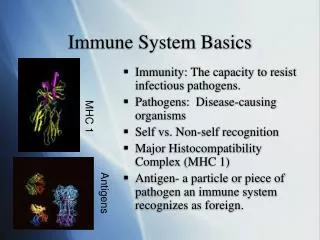
Immune System Basics. Immunity: The capacity to resist infectious pathogens. Pathogens: Disease-causing organisms Self vs. Non-self recognition Major Histocompatibility Complex (MHC 1) Antigen- a particle or piece of pathogen an immune system recognizes as foreign. MHC 1. Antigens.

E N D
Immune System Basics • Immunity: The capacity to resist infectious pathogens. • Pathogens: Disease-causing organisms • Self vs. Non-self recognition • Major Histocompatibility Complex (MHC 1) • Antigen- a particle or piece of pathogen an immune system recognizes as foreign. MHC 1 Antigens
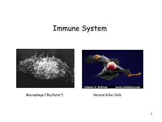
1st Defense:Non-specific Immune System • Reacts immediately after infection- does not need to ID pathogen. • Barrier Defenses: Skin and Mucous membranes • Inflammatory Defenses: • Histamine is released at the sign of damage • Blood vessels leak fluid and WBC’s • Cellular and Molecular Defenses: • Macrophages: Use pocket transport (phagocytosis) to destroy foreign particles. • Natural Killer Cells (NK): Release hydrolytic enzymes onto target cells to rupture/destroy them. • Interferon • Complement
Final Defense: Specific Immune System Recognizes pathogens and develops a sustained immune response. Comprised of two parts: • Cell- Mediated Response • Humoral Response White blood cells characters (lymphocytes): Macrophage Helper T cells (Th) Killer T cells (Tc) B cells
Specific Immunity- The Battle Begins! • Macrophages search body tissues for pathogens. • Consume pathogens with phagocytosis, kill it with lysosomes, and save the antigens. • Antigens placed into MHC2 receptors and displayed on macrophage’s membrane. • The macrophage is now considered an antigen-presenting cell (APC). • http://www.youtube.com/watch?v=eVLO6j6Ho64
Specific Immunity Cont. • Macrophage chemically signals Helper T to attach to it. • Helper T attaches to the MHC 2 receptor (with foreign antigen stuck in it) with its CD4 receptor. • Helper T cells have incredible variety of receptors that act like a “lock and key” in regards to the displayed antigen. • If the Helper T’s “key” fits the displayed antigen’s “lock”, the Helper T is activated. • Activation results in Helper T releasing cytokines (ex. Interleukin)- chemicals that cause lymphocytes to start mitosis.
Fig. 43-17 Antigen- presenting cell Peptide antigen Bacterium Class II MHC molecule CD4 TCR (T cell receptor) Helper T cell + Cytokines Humoral immunity (secretion of antibodies by plasma cells) + Cell-mediated immunity (attack on infected cells) + + B cell Cytotoxic T cell
Cell-Mediated ResponseSeek and Destroy • Body cells can be infected by viruses that will hide inside the cell. • As the virus reproduces inside cells, pieces of it fall off and are put into new MHC 1 receptors that the cell puts on its own membrane. • Killer T cells can bind to an infected cell’s MHC 1 receptors with their CD8 receptors. • If Killer T binds to MHC 1 receptors with antigen attached, it releases a chemical called perforin. • Perforin ruptures the infected cells membrane and exposes the virus to other immune cells. http://www.youtube.com/watch?v=1tBOmG0QMbA&feature=related
Fig. 43-11 Top view: binding surface exposed to antigen receptors Antigen Class I MHC molecule Antigen Plasma membrane of infected cell
Fig. 43-18-3 Released cytotoxic T cell Cytotoxic T cell Perforin Granzymes CD8 TCR Dying target cell Class I MHC molecule Pore Target cell Peptide antigen
Fig. 43-12 Microbe Antigen- presenting cell Infected cell Antigen associates with MHC molecule 1 Antigen fragment Antigen fragment 1 1 Class I MHC molecule Class II MHC molecule 2 2 T cell receptor T cell receptor 2 T cell recognizes combination (a) Cytotoxic T cell (b) Helper T cell
Humoral SystemBring in the artillery! • B cells have receptors called antibodies (100,000/cell). • Different B cells have uniquely shaped antibodies that match specific antigens. • If a B cell’s antibody is able to bind with a specific antigen (lock and key effect), the B cell receives a message from Helper T’s to become activated. • Activated B cells divide into Plasma B and Memory B cells. • http://www.youtube.com/watch?v=iDYL4x1Q6uU&feature=related
Humoral System Cont. • Plasma B cells produce and secrete 10,000 “keyed” antibodies per hour. • Due to their shape, each can bind to several antigens at once. • Antigen/Antibody binding has three effects. • Neutralization • Macrophage signaling • Complement pore formation • http://www.youtube.com/watch?v=lrYlZJiuf18&feature=fvw
Fig. 43-21 Viral neutralization Opsonization Activation of complement system and pore formation Bacterium Complement proteins Virus Formation of membrane attack complex Flow of water and ions Macrophage Pore Foreign cell
Memory B cells • These cells do not actively produce antibodies • Instead, they remain in the bloodstream and maintain their cell life cycle independently from Th commands. • If the same pathogen/antigen complex presents itself in the future, these cells are already activated and ready to produce antibodies. • There are also Memory versions of Th and Tc cells that serve a similar function.