Visual Fields
540 likes | 899 Views
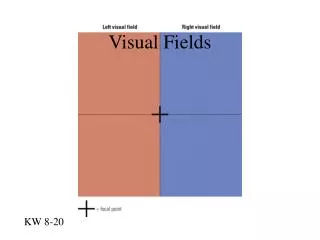
Visual Fields. Definition. Island of vision in sea of blindness. The peak of the island represents the point of highest acuity, the fovea while the “bottom less pit” represents the blind spot, the optic disc. Normal Monocular & Binocular Field. Monocular: Nasal side - 50° to 60°

Visual Fields
E N D
Presentation Transcript
Definition • Island of vision in sea of blindness. The peak of the island represents the point of highest acuity, the fovea while the “bottom less pit” represents the blind spot, the optic disc.
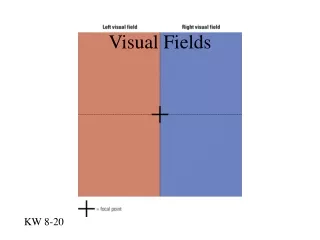
Normal Monocular & Binocular Field Monocular: • Nasal side - 50° to 60° • Superior side - 60° to 70° • Inferior side - 70° to 80° • Temporal side - 100°to110° Binocular: • Vertical field - 110° • Horizontal field - 200°
Visual Field Testing 1.Stimuli: Testing the island of vision at various levels requires targets that vary in (a) Size(b) Intensity(c) Colour 2. Field Test Methods: Kinetic; Mapping the contours of the island at different levels, resulting in one Isopter for each level tested. Static; Vertical contours of the island along a selected meridian.
Clinical testing methods • Central fields - Below 30° • Peripheral fields – Above 30° to 360° Central Fields: Confrontation method Amsler’s grid Bjerrum’s screen Peripheral Fields: Listen perimeter Goldmann perimeter
Defects • Retina • Glaucoma • Neuro
Retina Field defect • Central Scotoma: * Central serious Retinopathy * Macula Degeneration * Macular Oedema ( any macular disease) *Centro-cecal scotoma * Toxic Amblyopia * Stargadits
Field Defect • Contraction & Tubler field * Retinitis pigmentosa * High myopia * CRAO with Sparing of cilioretinal artery
Field Defect • Altitudinal defect: * AION • Depression: * Retinal Detachment • Ring Scotoma: * High myopia * Aphakic spectacle correction * Retinitis pigmentosa * Pan retinal photocoagulation
Glaucoma Field defect Isopter contraction: Peripheral Isopter contraction may be significantly smaller prior to any filed loss. Baring of the blind spot: It is also considered to be an early field defect in glaucoma.
Field defect • Angio-scotomata: Are long branching scotomata above or below the blind spot which are presumed to be resulted from shadow created by large retinal vessels and are felt to be an early change • Para central scotoma: One or more isolated Para central scotomata develop in the Bjerrum or arcute area.
Field defect • Seidel’s scotoma: A sickle shape defect arises from the blind spot and tapers to a point in a curved course with concavity towards the fixation point. • Bjerrum’s or Arcute scotoma: A relatively larger area of defect in the form of arching scotoma, which eventually fills the entire arcute area, from blind spot to the median raphe. With further progression, a double arcuate (ring or annular ) scotomata will develop.
Field defect • Roenne’s nasal step: The arcuate defects may not proceed at the same rate in the upper and lower portion of the eye. Consequently, a step-like defect is frequently created where the arcuate defects meet at the median raphe. This is called Roenne’s nasal step, and it is mostly superior nasal step as, Superior field is involve somewhat more frequently.
Field defect • Contraction & Tubler field: Generalized constriction of the peripheral field along with double arcuate scotoma- leads to Tubler field of vision in which only central vision remains clear.
Anatomy of the Visual Pathway • The Visual pathway (i) The end-organ –It is the neural epithelium of the rods & cones (ii) The first order neurone – It is bipolar cell with its axons in the inner layer of the retina. (iii) The second order neurone – It is ganglion cell of the retina its axons passes in to the nerve fibre layer and along the optic nerve to the LGB (iv) The third order nerurone – Originates in the cells of LGB, then travels by way of the optic radiations to the occipital cortex ( Visual centre)
The visual pathway thus consist of • Two Optic nerves • An Optic chiasma • Two Optic tracts • Two LGB • Two Optic radiation • Visual cortex on each side
Optic nerve • It extents from the lamina crib Rosa up to the optic chiasma • The total length of optic nerve- 3.5cm to5.5cm it can be divided into four parts: (i) Intra ocular part - 1mm (ii) Intra orbital part - 2 to 3cm (iii) Intracanalicular part - 4 to10mm (iv) Intracranial part - 1cm
Diseases of the Optic nerve • Congenital & Hereditary • Traumatic • Tumour • Inflammatory • Toxic • Vascular Lesion
Congenital • Mylinated nerve fiber defect • Coloboma • Hypoplasia • Drusen • Optic nerve pits • Tilted disc
Field defects • Mylinated nerve fiber *Blind spot enlargement *Paracecal scotoma *Ring scotoma *Central scotoma • Coloboma *Superior nasal depression *Contraction *Superior altitudinal hemianopia
Field defect • Hypoplasia: * Central Scotoma * Binasal&Bitemporal hemianopia * Bilateral inferior extension of blind spot • Drusen: * Blind spot enlargement * Irregular nerve fiber bundle scotoma
Field defect • Optic nerve pits: * Blind spot enlargement with or without macula involvement. * Central scotoma * Altitudinal hemianopia * Upper temporal field defect • Tilted disc: * Upper temporal defect which may be mistaken for chiasmal compression.
Hereditary Field defect • Optic atrophy (Lepers) * Contraction * Tubler Field • Retinitis Pigmentosa * Contraction * Ring scotoma * Tubler field
Traumatic Field defect • Traumatic optic neuropathy: * Superior altitudinal defect * Total Blind
Tumour Field defect • Glioma: * Blind spot enlargement * Contraction * Tubler field • Meningioma: * Junctional scotoma * Central scotoma * Upper temporal field defect
Inflammatory Field defect • Papillitis: * Central scotoma * Centro-cecal scotoma * Para central scotoma * Blind spot enlargement • Retro bulbar neuritis: * Central scotoma * Para central scotoma * Sectoral scotoma * Ring scotoma • Neuro retinitis: * Central Scotoma * Centro- cecal scotoma
Toxic Field defect • Mild Toxic : * Central scotoma * Centro- cecal scotoma (BE) • Severe Toxic : * Peripheral contraction * Total blind
Vascular lesionField defect • Anterior Ischemic optic neuropathy: * Altitudinal hemianopia ( mainly involving the inferior half)
Lesion due to pressure • Papilloedema: * Blind spot enlargement * Peripheral contraction * Total loss of visual field
ChiasmaField defect • Infra chiasmatic lesion: * Bitemporal hemianopia • Supra chiasmatic lesion: * Central hemianopic scotoma * Bitemporal hemianopia * Junctional scotoma * One eye is more field defect and other is less field defect (inferior temporal)
Field defect • Posterior side: * Infra Temporal field defect * Bilateral inferior quadrant scotoma ( near fixation point) * Bitemporal hemianopia
Optic Tract Field defect • Part I: * Homonymus hemianopia • Part II: * Incongruous hemianopia
LGB Field defect • Congruous Homonymus lower Quadrantropia • Congruous Homonymus upper Quadrantropia
Optic radiation Field defect • Bie in the Sky ( superior homonymus quadrantropia ) • Bie in the floor ( Inferior homonymus quadrantropia )
Visual Cortex Field defect • Congruous homonymus hemianopia • Congruous quadrantropia • Homonymus hemianopia with macula sparring • Homonymus hemianopia with macula splitting • Altitudinal hemianopia • Tubler field