Download
1 / 28
280 likes | 696 Views
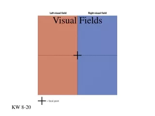
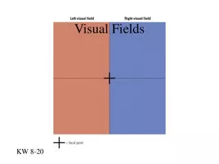
Visual Fields. KW 8-20. Fovea on Cortex. KW 8-22. Occipital Lobes are Independent. KW 8-24. Visual Areas. Areas V1 – V5. KW 8-17. Visual Cortex. Receptive Fields. Figure 6.16 Receptive fields

E N D
Visual Fields KW 8-20
Fovea on Cortex KW 8-22
Occipital Lobes are Independent KW 8-24
Areas V1 – V5 KW 8-17
Receptive Fields Figure 6.16 Receptive fields The receptive field of a receptor is simply the area of the visual field from which light strikes that receptor. For any other cell in the visual system, the receptive field is determined by which receptors connect to the cell in question.
Neural Activity KW 8-25
Overlap in Receptive Fields KW 8-27
Simple Cells 1 Mapping the Visual Cortex Hubel and Wiesel Nobel Prize Winners KW 8-29
Simple Cells 2 KW 8-29
Complex Cells KW 8-30
Neural Basis of Visual Perception The Cerebral Cortex: The Shape Pathway Hubel and Wiesel’s Cell Types in the Primary Visual Cortex Simple Cells Straight lines in particular orientation Complex Cells Straight lines in particular orientation in motion Hypercomplex cells Edge detector (edge in motion)
Hypercomlex Cell KW 8-31
Column Organization KW 8-34
Lesions in visual cortex KW 8-39
Dorsal and Ventral Streams Where What KW 8-14
Neural Basis of Perception Disorders of Object Recognition Visual Agnosia- Inability to Recognize Objects Prosopagnosia- Inability to recognize faces Contralateral Neglect - neglect left side of visual world
Injury to the “What” Pathway KW 8-40
Temporal Columns KW 8-35
Raderscheit Self-portraits Neglect 3.5 mo 2 mo 9 months after right parietal stroke 6 mo
Neglect Example 1 Left Visual Field Right Visual Right Brain Left Brain
Neglect Example 2 Left Visual Field Right Visual Right Brain Left Brain
Neglect Example 3 Left Visual Field Right Visual Right Brain Left Brain