Download
1 / 7
70 likes | 187 Views
Cardiovascular & Respiratory System . Chapter 37. Cardiovascular System. Heart – center of the CV system, hollow muscular organ made up of cardiac tissue, size of your fist, sits slightly to the L of midline

E N D
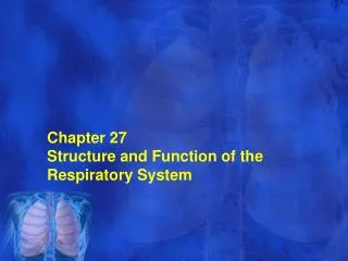
Cardiovascular & Respiratory System Chapter 37
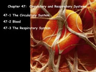
Cardiovascular System • Heart – center of the CV system, hollow muscular organ made up of cardiac tissue, size of your fist, sits slightly to the L of midline • Parietal pericardium or pericardial sac - loose fitting protective membrane around the heart, anchors heart • Walls of the Heart are composed of: • 1. Epicardium - pericardium • 2. Myocardium - cardiac muscle tissue • 3. Endocardium - lines the heart
Chambers • 2 Atria - upper 2 ventricles - lower • Arteries - thick muscular walls, carries blood away from the heart • Veins - thin walls with little muscle, blood towards heart and valves to prevent backflow • Capillaries - thin walled, single cell, connects arteries to veins • Superior vena cava & inferior vena cava bring blood to (R) atrium Coronary Veins- vein that brings blood from heart walls
Deoxygenated blood - blood to (R) atrium , (R) atrium contracts and blood goes into the (R) ventricle, Atrioventricular valve or AV - tricuspid • Next blood goes to the pulmonary trunk (R) & (L) • To the Pulmonary Artery *which carries blood to the lungs where it dumps the CO2 and gets O2 • 4 pulmonary veins that empty into the (L) atrium then to the (L) ventricle stronger, thicker walls, has to pump thousands of miles of vessels LAV valve is bicuspid • Semilunar valve - arteries that leave the heart
Pulmonary semilunar - pulmonary trunk (R) ventricle • Aortic semilunar - (L) ventricle to aorta • Conduction System – intrinsic regulating system, cells become specialized for impulse transmission • Node – compact mass of conducting cells • Sinoatrial or SA node (pacemaker) – ® atrial wall initiates rate or pace of heart ( can be altered by ANS, bloodborne chemicals, epinephrine, thyroid) • Impulse spreads over both atria causing contraction Stimulates AV node or Bundle of His – inter-ventricular septum • Purkinje fibers stimulate actual contraction of ventricles
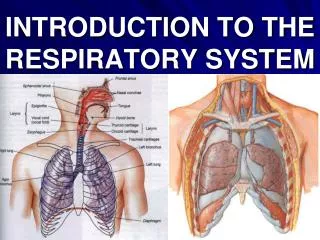
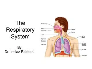
Gas Exchange • Respiration – process of exchanging O2 for CO2 • 1. Breathing – movement of air into and out of lungs • Inhale – diaphragm moves down, ribs up and out , cavity expands pressure in chest decreases, and air rushes in • Exhale – volume in chest cavity reduced, internal pressur greater so air is forced out • 2. Exchange of gases between internal surface of lungs and blood – diffusion between alveoli and capillary walls
3. Exchange of gases between blood and tissue cells • Heme – combines 4 O2 molecules gives it up by diffusion to tissues that need oxygen • Globin – carries the CO2 • Air exchange goes from nostril, nasal cavity, pharynx, larnyx, trachea, bronchus, lungs, where it exchanges with alveoli (300 million)