Download
1 / 17
170 likes | 290 Views
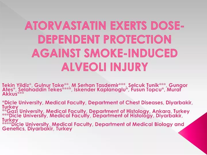
ATORVASTATIN EXERTS DOSE-DEPENDENT PROTECTION AGAINST SMOKE-INDUCED ALVEOLI INJURY. Tekin Yildiz *, Gulnur Take**, M Serhan Tasdemir ***, Selcuk Tunik ***, Gungor Ates *, Selahaddin Tekes ****, Iskender Kaplanoglu *, Fusun Topcu *, Murat Akkus ***

E N D
ATORVASTATIN EXERTS DOSE-DEPENDENT PROTECTION AGAINST SMOKE-INDUCED ALVEOLI INJURY TekinYildiz*, Gulnur Take**, M SerhanTasdemir***, SelcukTunik***, GungorAtes*, SelahaddinTekes****, IskenderKaplanoglu*, FusunTopcu*, Murat Akkus*** *Dicle University, Medical Faculty, Department of Chest Diseases, Diyarbakir, Turkey **Gazi University, Medical Faculty, Department of Histology, Ankara, Turkey ***Dicle University, Medical Faculty, Department of Histology, Diyarbakir, Turkey ****Dicle University, Medical Faculty, Department of Medical Biology and Genetics, Diyarbakir, Turkey
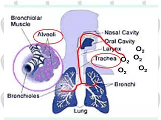
Objectives • We conducted this study to establish whether statins have protective anti-inflammatory effects against inflammation induced by cigarette smoke in rat lung cells, and in particular, the alveoli epithelial types I and II.
Materials and Methods • Thirty mature male Wistar albino rats were used in this experimental study. • The rats were divided into three groups of 10 rats each. • All rats were provided standard pelleted food and tap water ad libitum, and were kept at 20 ± 2ºC under a 12 h light/12 h dark light regimen in a stainless steel cage.
Materials and Methods • Thirty rats were divided into three groups and exposed to 20% smoke from 10 standard test cigarettes daily for 15 days. • The group-1 [G-1] smoke-exposed rats were used as controls, and • Groups 2 and 3 [G-2 and G-3] were treated with 0.5 mg/kg/day or 1.0 mg/kg/day atorvastatin, respectively. • The lung tissues were examined by transmission electron microscopy [TEM] [JEOL 1010, JEOL Ltd., Tokyo, Japan].
Materials and Methods • The study was approved by the institutional review board on animal experimentation of Dicle University, Diyarbakir, Turkey [2006-TF-059].
Procedure: • The rats were euthanized by ether anesthesia. • The lungs were extirpated and fixed in 2.5% phosphate-buffered glutaraldehyde for histopathological evaluation. • Samples of semi-thin cross sections of the lung tissues were prepared with an ultramicrotome, stained with toluidine blue, and stored in copper grids. • The tissues that were stained with lead acetate and uranyl citrate were screened by TEM [JEOL 1010, JEOL Ltd., Tokyo, Japan].
Figure 1a • G-1 [cigarette smoke control group; no atorvastatin] • Minimal intracytoplasmic edema [ICE] and partial separation were observed in the ATI nuclear membranes. • Mitochondrial crystolysis [MC] was detected in the capillary endothelial cells, while several pinocytic vesicles were seen in the capillary endothelial cells. • Furthermore, the rER tubulae of the capillary endothelial cells were of normal shape and size
Figure 1b • G-1 [cigarette smoke control group; no atorvastatin] • Microvillus deformation, pseudopod formation, edema, mitochondrial swelling, and MC were detected in the ATII cells. • All intracellular lipid droplets were clear, the mature lamellar body was absent, and the rERtubulae were inactive
Figure 2a • G-2 [0.5 mg/kg/day atorvastatin group] • No changes were detected in the G-2 ATI cells, blood–air barrier, or the cellular organelles. • The common basal membranes of the ATI and capillary endothelial cells were normal
Figure 2b • G-2 [0.5 mg/kg/day atorvastatin group] • Minimal increases in the collagen fibrils structure were observed in the interstitial areas
Figure 2c • G-2 [0.5 mg/kg/day atorvastatin group] • Upon examination of the ATI and ATII cells located beneath the collagen matrix, we detected only minimal changes in the mitochondriae and microvillae in the ATII cells. • The lysosomal structure was approximately normal-shaped, and minimal intracellular edema was detected in the ATI cells
Figure 3a • G-3 [1 mg/kg/day atorvastatin group] • Hyperplasia of the common basal membrane of the ATI and capillary endothelial cells was observed. • Hypertrophy, MC, and intracytoplamsic edema were detected in the ATI cells
Figure 3b • G-3 [1 mg/kg/day atorvastatin group] • A widespread increase of collagen fibrils was seen in the intra-alveolar septum
Figure 3c • G-3 [1 mg/kg/day atorvastatin group] • Chromatin condensation, atrophic appearance, cell shrinkage, and cytoplasmicvacuolizations were discerned in the ATII cells
Figure 3d • G-3 [1 mg/kg/day atorvastatin group] • The rER tubulae were spiral-shaped in the ATII cells [Figure 3d], closely resembling cells undergoing apoptosis.
CONCLUSIONS • The protective and/or hazardous effects of statins in lung cells may be dose-dependent. • Moreover, the administration of high dose atorvastatin can cause apoptosis in cells inflamed by cigarette smoke.