Download
1 / 25
270 likes | 320 Views
ECHOCARDIOGRAM (ECHO) FOR MITRIAL REGURGITATION. Dr C.S Mahlangu University of Limpopo(medunsa) Cardiothoracic registrar. Definition. Pathologic back flow of blood from the LV to the LA. Problem of leaflets,anulus or subvulvular apparatus. Modalities . Transthoracic echo.

E N D
ECHOCARDIOGRAM (ECHO) FOR MITRIAL REGURGITATION Dr C.S Mahlangu University of Limpopo(medunsa) Cardiothoracic registrar
Definition • Pathologic back flow of blood from the LV to the LA. • Problem of leaflets,anulus or subvulvular apparatus
Transthoracic echo PARASTERNAL LONG AXIS PARASTERNAL LONG AXIS PARASTERNAL LONG AXIS APICAL FOUR CHAMBER APICAL FOUR CHAMBER APICAL FOUR CHAMBER LV RV RA LA
Transthoracic echo SHORT AXIS LV AT PAPPILARY LEVEL RV RV IVS IVS AL LVD PM LVPW LVPW
TEE • Use of TEE • Pre op • Intra op • Indications • Limitations • Intra op • MR assessment • Assessment of repair
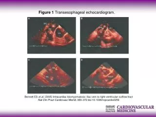
Monoplane transesophageal echocardiography LAA A1 Ao P1 A2 P2 A3 P3
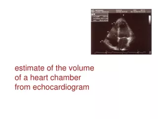
Anatomic echo parameters • LV dimension/size • LA dilation • LV volume • Stroke volume • Valve abnormality
LA SIZE APICAL 4 CHAMBER LV RV RA LA Normal size = 19-40mm
LA SIZE PARASTERNAL LONG AXIS RV RV AO LV IAS LAD LA LA wall
LV DIMENSIONS PARASTERNAL LONG AXIS RV IVS LV LVD LA NORMAL VALUES IVSd 6-12 LVEDD 36-52 LVPWd 6-11 IVSs 6-9 LVESD 24-42 LVPWs 9-14 LV EF 50-75% LVPW
Colour flow doppler • Jet area • Jet area index to LA • Vena contracta width RV LA RA
Central vs eccentric jet TRANSTHORACIC TRANSOESOPHAGEAL LV RV RA LA
Pulmonary flow reversal RV RA
Etiologies of mitral regurgitation • Normal leaflet motion : • annular dilatation (LV dilatation, ischemia) • leaflet perforation (trauma, endocarditis, ostiumprimum ASD) • Excess leaflet motion : • mitral valve prolapse (Barlow’s disease, Marfansyndrom, Ehler-Danlossyndrom, pseudoxanthomaelasticum) • chordae rupture (prolapse, trauma, endocarditis) • papillary muscle rupture (infarction, trauma) • Restricted leaflet motion : • leaflet retraction (rheumatic fever, carcinoïd heart diasease, systemic lupus, annular calcifications, ...) • systolic anterior motion (HCOM) • chordae retraction (rheumatic disease) • papillary muscle retraction and/or dysfunction (aneurysm, infarction, ischemia, endomyocardial fibrosis)
Various forms of mitral regurgitation Mitral prolapse in Barlow’s disease Mitral prolapse with ruptured chords Infective endocarditis Rheumatic disease
Rheumatic RV LVOT LV LA
Prolapse and flail leaflet MYXOMATOUS BARLOW’S DISEASE ISCHEMIC TRAUMA ENDOCARDITIS
Annular dilatation PARASTERNAL LONG AXIS APICAL 4 CHAMBER RV LV LV LV LA RA LA


![DOWNLOAD [PDF EPUB] Echo North By Joanna Ruth Meyer [EBOOK KINDLE]](https://cdn4.slideserve.com/8150584/slide1-dt.jpg)