Download

1 / 33
400 likes | 1.39k Views
Urinalysis 101. Dr. Bryon Hargrove Fall 2012. Purpose of Urinalysis. Urine analysis is considered to be the oldest clinical chemistry test . At present, it still remains one of the basic tests regardless of the disease involved.

E N D
Urinalysis 101 Dr. Bryon Hargrove Fall 2012
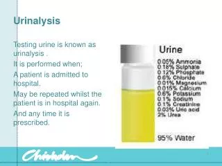
Purpose of Urinalysis • Urine analysis is considered to be the oldest clinical chemistry test. • At present, it still remains one of the basic tests regardless of the disease involved. • Currently, chemical testing of urine is usually performed by use of a test strip. • The routine urine examination includes test strip screening, followed by microscopic examination of urinary sediment if necessary. • Other tests are performed where applicable. (100 Tests)
Urine Sampling • A midstream urine specimen collected after thorough hygiene of external genitals is an optimal sample. • Appropriate urine sampling and preparation are of utmost importance for the chemical and microscopic urine tests to produce reliable results. • Therefore, the containers used for urine collection must be thoroughly clean and time of analysis strictly defined (within 1-2 hours from urine sampling). • Urine specimen can be obtained by: • spontaneous urination • spontaneous urination - midstream urine specimen • catheterization • percutaneous and suprapubic urinary bladder aspiration.
Types of Urine Specimen • A healthy adult excretes about 1500 mL of urine daily. The overall amount of urine excreted during 24 hours should preferably be obtained for urinalysis, be it quantitative or qualitative. • However, this mode of urine collection is quite questionable, because not all people comply with the instructions and may not present total 24-hour urine for examination. • Urine is collected into clean containers in three ways: • random specimen (individual specimen) • diurnal or nocturnal specimen collected over a certain period of time • 24-hour urine • For routine urine examination, itisrecommended to use the first morning urine obtainedat least 8 hoursfrom the last void. • For urinalysis by means of test strip, anyrandom urine specimencanbeused, however, the time of urine samplingshouldbeconsidered on resultinterpretation.
Random Specimen • This type of urine specimencanbeobtainedatany time of the day, however, the first morning urine isrecommended as itismostconcentrated and approximates the composition of 24-hour urine. • The physicochemical examination of urine isperformed on fresh urine without the addition of preservative.
Diurnal or Nocturnal Urine Specimen • The urine collected in thiswayisused for urinalysis in somedisorders, e.g. alimentaryglycosuria, proteinuria, etc. The mode of urine collection, storage and preservationdepends on the tests to beperformed.
24 Hour Urine Sample • For quantitative assays, 24-hour urine specimensshouldbecarefullycollected and preferablyrefrigerated. • Certain preservativesshouldbeadded if chemically instable compounds are to bedetermined. • The type of preservativedepends on the analyte to bedetermined. 10% solution of thymol in isopropanol (5 ml for 24-hour urine) ismostcommonlyused as preservative.
Containers • The containers used for urine collection must be made from a material non-reactive with urine constituents. They must have a wide neck (at least 4 cm in diameter), and be 50 ml in size, preferably disposable. The container used for urine collection must be thoroughly clean. • In case of urine specimen transportation, the containers must be provided with appropriate caps and labels containing the following data: • patient's first and last name, • date and time of urine sampling, and • sample storage before transportation to laboratory (in a refrigerator at +2 °C to +8 °C or at room temperature).
Storage of Urine Specimen • The analysis of a urine specimen should be carried out as soon as possible after urine sampling, due to rapid autolytic changes of the formed elements (cells, cylinders) and propagation of bacteria. Therefore, urine specimens should not be stored at room temperature for more than 2 hours (according to some sources, not more than 30 minutes). It is recommended for urine specimen to be stored at +2 °C to +8 °C, however, the time of storage should be maximally reduced. • If stored in a refrigerator, urinalysis should be carried out within 4 hours from urine sampling
Consequences of Inappropriateurine specimen storage • Until analysis, urine should be protected from direct sunlight, thus avoiding the possibility of erroneous results due to subsequent urine changes. In case of prolonged urine storage beyond the above mentioned time limits, the following changes develop: • bilirubin- decreased due to sun exposure • color- changed due to oxidation or metabolite reduction • bacterial count– increased • cell and cylinder count - decreased due to disintegration, especially in a diluted or alkaline specimen • glucose- decreased due to glycolysis and bacterial utilization • ketones- decreased due to volatilization • nitrites- increased due to bacterial reduction of nitrates • pH- increased because of urea decomposition to ammonia due to the bacterial urease activities • turbidity- increased due to bacterial growth and possible deposition of amorphous salts • urobilinogen- decreased due to oxidation to urobilin
Procedure of urine sampling: Urine Collection • Urine specimen • Morning urine (midstream) • Patient preparation • The patient is given a urine container, labeled with the patient's name and date • The patient is instructed on how to perform urine sampling: • hand wash with soap • washing external genitals with soap • genitals hygiene • the first void should be allowed to pass into the lavatory (1) • the midstream urine is voided into the urine container (2) • then the remaining urine is voided into the lavatory (3) • the container is closed and urine specimen immediately delivered to the laboratory.
Men versus Woman: Avoid Contamination • Men • Uncircumcised men should be warned to pull the preputium to expose the external ureteral orifice. • The glans should be cleaned with a clean (sterile) towel, from the ureteral orifice outward. • Women • The ureteral orifice and surrounding area should be cleaned with a clean (sterile) towel.
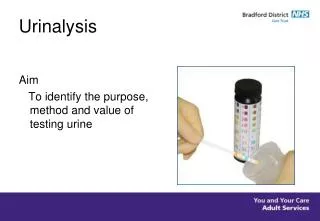
Test Strips (FYI) • Test strips are used to analyze urine pH and specific gravity, and to demonstrate the presence of glucose, bilirubin, ketones (acetoacetic acid), blood, protein, urobilinogen, nitrites, leukocytes and ascorbic acid in urine (depending on the type of test strip used). • Test strips are used for rapid and reliable demonstration and semiquantitative determination of particular analytes in urine by quite a simple procedure. • While working in a medical biochemistry laboratory in Vienna in 1920s, Fritz Feigl began to think about a more rapid testing for certain urine compounds by filter paper impregnation with particular reagents. Some 20 years elapsed from the idea to its implementation. In 1935, a report on rapid urinalysis by test strips appeared in the journal of the Leipzig Academy of Sciences. • However, Feigl's invention found routine application years later, when industry manufacture of strips for rapid urine testing started. First strips were intended for a single parameter, e.g. glucose, albumins, ketones, etc. • Then, Ames started the manufacture of test strips for simultaneous determination of a number of analytes, e.g. Hema-combistix for protein, glucose, pH and acetone. • The shortcoming of these first test strips was their instability due to the reagent susceptibility to the effect of moisture (with filter paper as a medium). Later on, filter paper as a carrier was substituted by inert plastic strips with reagent-impregnated paper slips (6x6 mm slips of filter paper) pasted on. • Currently used test strips are made of plastic foil with reagent paper (impregnated reagents) at particular areas. A special layer to absorb excess fluid is placed underneath the reagent paper. A thin nylon mesh is stretched over the absorbent layer and reagent paper, fixing them to the foil and at the same time protecting them from any undesirable contact and contamination.
History of the Test Strips • The strip allows the reagent paper (6x6 mm) to be uniformly perfused with urine while the reaction is proceeding steadily, together with removal of excess urine. • The principles of reagent reactions in dry state for testing particular analytes in urine are identical to those performed in the test tube. • A specific chemical reaction for determination of an analyte concentration is combined with an indicator or conjugated reagent to produce a color of varying intensity proportional to the tested analyte concentration. • The first test strips were manufactured in 1950 for diagnostic purpose, i.e. for the detection and follow-up of diabetes mellitus. • These test strips contained reagents for demonstration of glucose in urine (Clinistix, Ames; S-Glucotest, Boehringer-Mannheim). Reagents for other tests were then gradually added onto the test strip.
What do the Test Strip test? • At present, 11 different urine parameters can be qualitatively determined by manual evaluation or automatically: • pH • Glucose • Protein • Nitrites • Urobilinogen • Ketones • Bilirubin • Blood • Leukocytes • Specific gravity • Ascorbic acid • http://www.drugcheck.com/hc_uc-healthscreen-10.html
Advantages and Disadvantage of Test Strips • Advantages: • Test strips allow rapid and reliable demonstration and semiquantitative determination of particular analytes in urine. • simple use and easy to use for quick results constant reagent composition • specificity for particular parameters, and long-term stability. • Disadvantages: • inadequate sensitivity for some analytes (e.g. albumin) • Human error in reading the results • susceptibility to interferences, andsemiquantitativeresults
Interpretation of Results http://faculty.washington.edu/kepeter/119/images/urinalysis_tests.htm
Color of Urine • Many things affect urine color, including fluid balance, diet, medicines, and diseases. • How dark or light the color is tells you how much water is in it. • Vitamin B supplements can turn urine bright yellow. • Some medicines, blackberries, beets, rhubarb, or blood in the urine can turn urine red-brown.
Clarity of Urine • Urine is normally clear. • Bacteria, blood, sperm, crystals, or mucus can make urine look cloudy.
Urine Odor • Urine does not smell very strong, but has a slightly "nutty" odor. • Some diseases cause a change in the odor of urine. • For example, an infection with E. coli bacteria can cause a bad odor, while diabetes or starvation can cause a sweet, fruity odor.
pH of Urine • http://www.rnceus.com/ua/uaph.html • Normal: 4.6-8.0 • The pH is a measure of how acidic or alkaline (basic) the urine is. • A urine pH of 4 is strongly acidic, 7 is neutral (neither acidic nor alkaline), and 9 is strongly alkaline. • Sometimes the pH of urine is affected by certain treatments. For example, your doctor may instruct you how to keep your urine either acidic or alkaline to prevent some types of kidney stones from forming. • Some foods (such as citrus fruit and dairy products) and medicines (such as antacids) can affect urine pH. • A high (alkaline) pH can be caused by severe vomiting, a kidney disease, some urinary tract infections, and asthma. • A low (acidic) pH may be caused by severe lung disease (emphysema), uncontrolled diabetes, aspirin overdose, severe diarrhea, dehydration, starvation, drinking too much alcohol, or drinking antifreeze (ethylene glycol).
Glucose in Urine • Glucose is the type of sugar found in blood. • Normally there is very little or no glucose in urine. • When the blood sugar level is very high, as in uncontrolled diabetes, the sugar spills over into the urine. • Glucose can also be found in urine when the kidneys are damaged or diseased. • Too much glucose in the urine may be caused by uncontrolled diabetes, an adrenal gland problem, liver damage, brain injury, certain types of poisoning, and some types of kidney diseases. • Healthy pregnant women can have glucose in their urine, which is normal during pregnancy.
Protein in Urine • Protein is normally not found in the urine. • Fever, hard exercise, pregnancy, and some diseases, especially kidney disease, may cause protein to be in the urine. • Bence Jones Protein • This is an abnormal protein found in the urine of about 50% of people with a rare type of cancer called multiple myeloma. • A urine test is often done when multiple myeloma is suspected. • Not normally performed in a UA
Nitrates in Urine • Bacteria (E. Coli) that cause a urinary tract infection (UTI) make an enzyme that changes urinary nitrates to nitrites. • Nitrites in urine show a UTI is present.
Ascorbic Acid • High levels of of Vitamin C can cause false positive tests. • High levels may or may not indicate contamination of the specimen • Could be an indicator for possible pregnancy if female.
Ketones • When fat is broken down for energy, the body makes substances called ketones (or ketone bodies). • These are passed in the urine. • Large amounts of ketones in the urine may mean a very serious condition, diabetic ketoacidosis, is present. • A diet low in sugars and starches (carbohydrates), starvation, or severe vomiting may also cause ketones to be in the urine. • Ketones in the urine can mean uncontrolled diabetes, a very low-carbohydrate diet, starvation or eating disorders (such as anorexia nervosa or bulimia), alcoholism, or poisoning from drinking rubbing alcohol (isopropanol). • Ketones are often found in the urine when a person does not eat (fasts) for 18 hours or longer. This may occur when a person is sick and cannot eat or vomits for several days. • Low levels of ketones are sometimes found in the urine of healthy pregnant women.
Bilirubin • This is a substance formed by the breakdown of red blood cells. It is passed from the body in stool. • Bilirubin is not found in urine. • If it is present, it often means the liver is damaged or that the flow of bile from the gallbladder is blocked.
Urobilinogen • This is a substance formed by the breakdown of bilirubin. • It is also passed from the body in stool. • Only small amounts of urobilinogen are found in urine. • Urobilinogenin urine can be a sign of liver disease (cirrhosis, hepatitis) that the flow of bile from the gallbladder is blocked.
Blood • Blood cells are not found in urine normally. • Inflammation, disease, or injury to the kidneys, ureters, bladder, or urethra can cause blood in urine. • Strenuous exercise, such as running a marathon, can also cause blood in the urine. • White blood cells may be a sign of infection or kidney disease.
Leukocytes • Leukocyte esterase shows leukocytes (white blood cells [WBCs]) in the urine. • WBCs in the urine may mean a UTI is present or inflammation of the tissues in the urethra or bladder. • Early Sign of Kidney Infection
Specific Gravity • Normal: 1.005-1.030 • This checks the amount of substances in the urine. • It also shows how well the kidneys balance the amount of water in urine. • The higher the specific gravity, the more solid material is in the urine. • When you drink a lot of fluid, your kidneys make urine with a high amount of water in it which has a low specific gravity. • When you do not drink fluids, your kidneys make urine with a small amount of water in it which has a high specific gravity. • A very high specific gravity means very concentrated urine, which may be caused by not drinking enough fluid, loss of too much fluid (excessive vomiting, sweating, or diarrhea), or substances (such as sugar or protein) in the urine. • Very low specific gravity means dilute urine, which may be caused by drinking too much fluid, severe kidney disease, or the use of diuretics.
Microscopic Analysis • In this test, urine is spun in a special machine (centrifuge) so the solid materials (sediment) settle at the bottom. • The sediment is spread on a slide and looked at under a microscope.
Results of Microscopic Analysis • Red or white blood cells. Blood cells are not found in urine normally. Inflammation, disease, or injury to the kidneys, ureters, bladder or urethra can cause blood in the urine. • Strenuous exercise, such as running a marathon, can also cause blood in the urine. • White blood cells may be a sign of infection or kidney disease. • Casts. Some types of kidney disease can cause plugs of material (called casts) to form in tiny tubes in the kidneys. The casts then get flushed out in the urine. Casts can be made of red or white blood cells, waxy or fatty substances, or protein. The type of cast in the urine can help show what type of kidney disease may be present. • Crystals. Healthy people often have only a few crystals in their urine. A large number of crystals, or certain types of crystals, may mean kidney stones are present or there is a problem with how the body is using food (metabolism) • Bacteria, yeast cells, or parasites. There are no bacteria, yeast cells, or parasites in urine normally. If these are present, it can mean you have an infection. • Squamous cells. The presence of squamous cells may mean that the sample is not as pure as it needs to be. These cells do not mean there is a medical problem, but your doctor may ask that you give another urine sample.