Download

1 / 49
600 likes | 1.14k Views
Urinalysis. Dr. A. Basu MD. Topic. Definition and normal urine Indication Collection Examination Physical Chemical Microscopical . Urinalysis : Definition. Qualitative and semi quantitative evaluation of renal products. Normal Urine = At a glance.

E N D
Urinalysis Dr. A. Basu MD
Topic • Definition and normal urine • Indication • Collection • Examination • Physical • Chemical • Microscopical
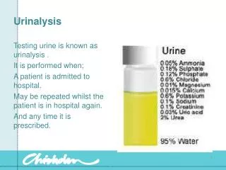
Urinalysis : Definition • Qualitative and semi quantitative evaluation of renal products.
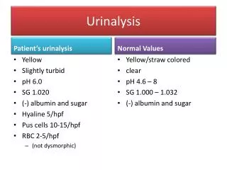
Normal Urine = At a glance • a. Physical - color - normally pale yellow • transparency - clear • specific gravity - 1.020 - 1.030 • b. Chemical - pH • acid in carnivores • alkaline in herbivores • protein - normally 0 mg/dl • glucose - normally none • blood - normally none • c. Sediment - urine solids stained after centrifugation( no finding, except scanty epithelial cell)
Indication • Glomerulonephritis • Diabetes mellitus • Urinary tract infection • Tumors
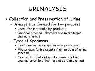
Collection • Random collection: Early morning: chemical are present in higher concentration • 24 hours urine collection Nephrotic syndrome • Suprapubic Trans- abdominal needle aspiration of the bladder (purest sample)
Urine preservatives • Boric acid • HCl • Sulfuric acid
Collection • Maintain patient privacy • Do not clean the area with antiseptic • Collect mid stream urine
When you should examine • Immediate • Or, within One hour after collection of urine.
The interval of time which elapses from collection to examination in the laboratory is important. Changes which occur with time after collection include: loss of ketone bodies, loss of bilirubin, overgrowth of contaminating microorganisms.
URINALYSISThree phases • A. Physical Examination • B. Chemical Examination • C. Microscopical Examination
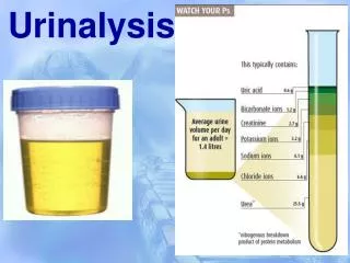
URINALYSIS A. Physical Examination • 1.Volume • 2. Color • Odor • 3.Appearance(e.g. transparent or Turbidity present) • 4.pH • 5. Specific Gravity
Colour • Red: Hematuria, hemoglobinuria, certain drugs, myoglobinuria . • White: chyluria • Red colour on standing : Porphyria • urine darkens on standing - Alkaptonuria
Odor • Normal: ammonia • In infection: fishy smell • If Ketone is high: sweet smell.
Appearance • Normal: transparent • Turbid: • High WBC • High epithelial cells.
ph • The glomerular filtrate of blood plasma is usually acidified by renal tubules and collecting ducts and from a pH of 7.4 to about 6 in the final urine.
Specific Gravity • Low (1.007 to 1.010) : End stage kidney disease. • High (1.035) : SIADH, contamination , presence of glucose.
Chemical test • Protein • Sugar (Glucose) • Ketone • Blood • Nitrite • Leukocyte Esterase
Protenuria • More than 150 mg/day is defined as proteinuria
Ketone • Diabetic ketosis or • Some other form of calorie deprivation (starvation).
Nitrite • Positive nitrite test ; Bacteria may be present
Leukocyte Esterase • Positive: WBC are present
URINE DIPSTICK CHEMICAL ANALYSIS • A dipstick is a paper strip with patches impregnated with chemicals that undergo a color change when certain constituents of the urine are present or in a certain concentration. • The strip is dipped into the urine sample, and after the appropriate number of seconds, the color change is compared to a standard chart to determine the findings
In protenuria 4+ Leucocyte esterase Colors' are before examination Nitrite • A sample Strip • For Chemical • Analysis Of • Urine pH Protein Blood Specific Gravity Ketone Glucose Bilirubin
Microscopical examination • RBC • WBC • Yeast • Casts • Crystals
Examination(Microscopy)RBC • Red blood cells in urine appear as refractile disks. • With hypertonicity of the urine, the RBC's begin to have a crenated appearance.
Examination(Microscopy)RBC • Glomerular damage • Tumors which erode the urinary tract anywhere along its length, • Kidney trauma, urinary tract stones, • Renal infarcts, acute tubular necrosis, • Upper and lower urinary tract infections, nephrotoxins, and physical stress. • Red cells may also contaminate the urine from the vagina in menstruating women or • From trauma produced by bladder catherization.
The presence of dysmorphic RBC's in urine suggests a glomerular disease such as a glomerulonephritis. Normal RBC
White Blood Cells : When we see this cell? • Infection (pyuria) • Acute glomerulonephritis
These white blood cells in urine have lobed nuclei and refractile cytoplasmic granules.
A white blood cell is seen at the left and a red blood cell at the right for size and morphologic comparison
Important • Tuberculosis may be a cause for a 'sterile pyuria' in which routine cultures fail to grow bacterial organisms, but inflammation is present.
Epithelial Cells • Acute tubular necrosis • UTI
Oval Fat Bodies • When lipiduria occurs, these cells contain endogenous fats. When filled with numerous fat droplets, such cells are called oval fat bodies
Oval fat bodies consist of degenerated tubular cells containing abundant lipid, which appears refractile
Casts • Hyaline casts are composed primarily of a mucoprotein (Tamm-Horsfall protein) secreted by tubule cells.
The presence of this red blood cell cast in on urine microscopic analysis suggests a glomerular or renal tubular injury.
This white blood cell cast suggests an acute pyelonephritis.
This renal tubular cell cast suggests injury to the tubular epithelium(ATN).
Presence of Granular cast suggest Chronic disease: chronic GN
Granular cast later form a finely granular cast, and ultimately, a waxy cast. Waxy cast. Note that the edges are sharp and there are "cracks" in this cast.
Yeast • Yeast cells may be contaminants or represent a true yeast infection. • Most often they are Candida, distinguished by their tendency to bud
Crystals MAY BE ASSOCIATE WITH STONE IN THE Urinary system.
These are oxalate crystals, which look like little envelopes (or tetrahedrons, depending upon your point of view).
These "triple phosphate" crystals look like rectangles, or coffin lids.