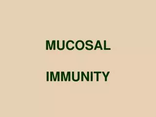
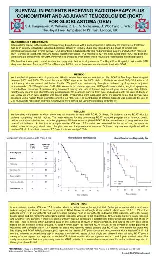
Download
1 / 31
440 likes | 1.85k Views
Endoscopic Mucosal Resection. Dr. Howard Mertz Clinical Assistant Professor Vanderbilt University Saint Thomas Hospital Nashville TN. Acknowledgements. Wilson Cook support for this presentation Olympus support for EMR training. Background:.

E N D
Endoscopic Mucosal Resection Dr. Howard Mertz Clinical Assistant Professor Vanderbilt University Saint Thomas HospitalNashville TN
Acknowledgements • Wilson Cook support for this presentation • Olympus support for EMR training
Background: • Endoscopic removal of superficial lesions in the GI tract feasible • This allows full pathologic evaluation superior to surface biopsies • Can be curative • Can prevent surgery
Background: • Endoscopic Mucosal Resection (EMR) now done more widely and safely • Targets • Large sessile colon polyps • Esophageal dysplasia or early cancers • Gastric cancers or benign tumor nodules • Duodenal polyps
5 Layers of the GI tract by EUS EUS Histology Mucosa MM SubMuc MP Serosa
Submucosal (SM) Invasion • Increases risk of lymph node metastases • Esoph Ca: sm1 8-30%, sm2 23%, sm3 44% • Gastric Ca: SM 2-25% • Colon Ca: SM 10-18% • If definite and more than superficially into SM layer by EUS, avoid EMR • If SM on path: surgery or Chemo/RT
Patient Selection and EUS: • EUS to evaluate depth except in polyps • Avoid EMR if submucosal cancer • No lymphadenopathy • Benign lesions deep in the submucosa • Avoid if previous snaring that will tether lesion down with scar tissue
T1, N0 Rectal Cancer Mass confined To mucosal layer Can be resected Transanal or by EMR
Nodule in Barretts Esophagus T1-2 N1
Mucosal Lesion Evaluation sm> mp> 53 yo man with heartburn and nodule in Barretts epithelium. EUS: mucosal/submucosal lesion
Submucosal Injection: • Create fluid cushion in submucosa • Protects muscularis propria from perforation • Volumes between 5 and 20 cc • Use Sclerotherapy needle • Injection fluids can be normal or hypertonic saline, D50, Hyaluronic acid • Methylene blue and epinephrine helpful
Submucosal Injection: • Normal Saline 18.5 cc • Epinephrine (1:10,000) 1 cc • Methylene blue 0.5 cc • If gastric, use D50 or methyl cellulose, due to faster diffusion Haber, Lennox Hill NY
Submucosal Injection: • Start on distal side of lesion • Inject several location • Look for lift up of lesion over cushion • Failure to lift indicates deeper penetration, contraindication to EMR • Methylene blue shows the cushion
Marking Tips • Mark lesion with burns from needle knife or polypectomy snare tip or APC • Can use indigo carmine or other dyes • Inject enough so cushion extends well beyond markings • Snare halfway up cushion
Techniques • Inject and snare • Inject, band and snare • Inject, suction cap, snare
Devices • Injection needle • Stiff snares: Hex snare best, braided helpful • Combined needle-snare (US Endo I snare) • Cap EMR on EGD scopes • Olympus EMR kit—largest, angled or straight • Cook Duett—variceal type bander, smaller • Roth net for retrieval of specimens
Mucosal Lesion Evaluation sm> mp> 53 yo man with heartburn and nodule in Barretts epithelium. EUS: mucosal/submucosal lesion
Endoscopic Mucosal Resection Submucosal Elevation Banding Snare Injection Resection Pathology: inflammatory polyp in Barretts
How to Ensure Successful EMR • Case selection: avoid non-lifting, difficult to access, near circumfrential disease • Can be more aggressive in rectum • Attempt en bloc resection when possible • Carefully resect, biopsy, burn residual • Close follow up < 6 months to recheck site • Discuss option of surgery
Risk of Perforation • Highest • Duodenum • Colon, Esophagus • Stomach • Rectum • Lowest • Reported Rates 0.1-5%
How to minimize Perforation • Avoid hot biopsy forceps if possible • Ensure good mucosal lift before snaring • Reinject saline if EMR taking more time and cushion diffusing out • Lift with snare prior to cauterizing
Bleeding Risk • Size < 1cm 0% • Size 1-2 cm 4% • Size 2-3 cm 24% • Size >3 cm 32% • By Site: Esophagus 11%, Stomach 28%, Duodenum 33%, Colon 17%
How to Minimize Bleeding • Slow steady closure of snare during cautery • Blended current or all coag • Argon laser to cauterize and bleeders • No anti-coagulants or NSAIDS for 2 weeks • May avoid epi to allow any bleeding to be overt initially
Summary • EMR is available and feasible • Requires expertise, EUS helpful • Complications include perforation (approx 2%) and bleeding (approx 6%) • Curative if mucosal disease only • Can prevent surgery