Download
1 / 38
450 likes | 868 Views
Celiac Disease and Gluten Sensitivity. A Case-based Approach to Gastroenterology. Kimberly Carter, MS, PA-C Division of Gastroenterology University of Pennsylvania Kimberly.Carter2 @uphs.upenn.edu. My bloating got better when I started a GFD…. Does that mean I have celiac disease?.

E N D
Celiac Disease and Gluten Sensitivity A Case-based Approach to Gastroenterology Kimberly Carter, MS, PA-C Division of Gastroenterology University of Pennsylvania Kimberly.Carter2@uphs.upenn.edu
My bloating got better when I started a GFD… Does that mean I have celiac disease?
Why Differentiate? • Patient • Improve well-being • Decrease intestinal symptoms • Reduce systemic complications • Practitioner • Risk Stratify • Screen family members • Healthcare Economics • Avoid unnecessary invasive and costly testing
Objective • Compare Celiac Disease (CD) vs. Gluten Sensitivity (GS) as it relates to serological/HLA testing and diagnostic work-up • NOTE: No diagnostic criteria for non-celiac gluten sensitivity • Review diagnostic algorithm of CD • Identify limitations of serologic testing • Understand the utility of HLA testing • Define gluten sensitivity (GS) • Discuss management of CD and GS
Case Study # 1 23-year-old female with Type I DM presents with a 1 year history of abdominal cramping accompanied by bloating, gas, and alternating constipation and diarrhea in the setting of a 10 lbs. weight loss.
Labs • Immunoglobulin A 243 (50-500 mg/dL) • Tissue Transglutaminase IgA 58 (H) <=19 unit(s) • TSH 2.90 (0.27-4.20 uIU/mL) • Hemoglobin 9.5 (L) (12.0-16.0 g/dL) • Hematocrit 30 (L) (36-46 %) • MCV 71 (L) (80-100 fL) • Ferritin 5 (L) (13-150 ng/mL) • Iron 16 (L) (28-170 ug/dL)
Scalloped mucosa Duodenal mucosa with expansion of the lamina propria, increased intraepithelial lymphocytes and villous blunting
Case Review Young female with an elevated tTG IgA in the setting of luminal symptoms, weight loss, anemia, and diabetes with duodenal biopsies consistent with villous atrophy confirming celiac disease.
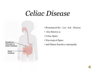
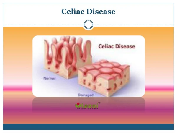
Celiac Disease • Chronic autoimmune disease of the small intestine triggered by the ingestion of gluten • Causes intestinal inflammation • Impairs absorption of nutrients • Contributes to systemic complications Milito T, Muri M, Oakes J, et al. Celiac disease: Early diagnosis leads to the best possible outcome. Journal of the American Academy of Physician Assistants. 2012;25(11):43-47.
Celiac Disease Celiac Disease Foundation
Establishing a diagnosis • Clinical suspicion • Serology • Biopsy • Therapy Response
Who should be tested • High risk groups • 1st degree relative • Type I Diabetes and Thyroid Disease • Down syndrome, Turner syndrome • Gastrointestinal symptoms • Misdiagnosed IBS/lactose intolerance • Asymptomatic/Extraintestinal manifestations
Extraintestinal manifestations • Osteopenia, osteoporosis • Reproductive disorders • Neuropsychiatric symptoms • Dermatitis herpetiformis • Nutrient Deficiencies • Elevated LFTs
Serologic testing • Appropriate initial diagnostic work-up and assess therapy response • Serology obtained on gluten containing diet • Immunoglobulin A (IgA) anti-tissue transglutaminase (tTG) • IgA endomysial antibody • IgG or IgA deamidated gliadin peptides (DGPs) • Quantitative IgA • 5% IgA deficiency .
Serologic diagnostic accuracy Fasano A, Catassi C. Celiac Disease. The New England Journal of Medicine. 2012;367:2419-2426.
Endoscopic evaluation • Gross Findings • Scalloping • Fold flattening • Fissuring • Nodular mucosa • Histologic features • Intraepithelial lymphocytes • Crypt hyperplasia • Villous blunting/atrophy (Marsh III Criteria) • NOTE: Absence of visual endoscopic findings does not exclude the disease Setty M, Hormaza L, Guandalini S. Celiac Disease Risk Assessment, Diagnosis, and Monitoring. Molecular Diagnosis & Therapy. 2008;12(5):289-298.
Management of Celiac Disease • Milito T, Muri M, Oakes J, et al. Celiac disease: Early diagnosis leads to the best possible outcome. Journal of the American Academy of Physician Assistants. 2012;25(11):43-47.
Gluten-free diet • Eliminates wheat, rye, and barley • Rice, corn, millet, potato, buckwheat, and soybeans are safe • Common gluten free foods • fresh fish, meats, milk, cheese, fruits, vegetables • Gluten-free substitutes are often expensive and may be difficult to access
Management of Celiac Disease • Annual Labs • CBC, CMP, anti-gliadin, tTG • Screen for and replete micronutrient deficiencies (iron studies, B1, B6, folate, B 12, Zinc) • Fertility counseling • Screen for osteopenia/osteoporosis with DEXA at diagnosis
Therapy Response • Clinical remission: Immediate • Serologic response: Weeks-months • Mucosal healing: 6-24 months • Poor response to GFD
Case Study # 2 • 26-year-old female with no significant PMH presents with 3 year history of abdominal discomfort accompanied by bloating, gas, and constipation. Symptom improvement on GFD. • ROS: headaches, fatigue and 15 lbs weight loss in the past 6 months. • Serology • Anti-endomysial <1:10 • tTG IgA < 5 • GliadinIgG 37 (H) • Gliadin IgA <20 • IgE 14.3 • Allergens: below detectable limits wheat
Diagnostic Dilemma • Suggestive clinical features but negative serologic tests • Gluten free diet • Selective IgA deficiency • Wheat allergy or gluten sensitivity • Seronegative celiac disease Kelly, CP. Diagnosis of celiac disease. In: UpToDate, Lamont, JT (Ed), UpToDate, Waltham, MA. (Accessed on April 30, 2014).
Diagnostic Dilemma • Positive serologic tests but negative small bowel biopsies • False positive serology • 1st generation gliadin Kelly, CP. Diagnosis of celiac disease. In: UpToDate, Lamont, JT (Ed), UpToDate, Waltham, MA. (Accessed on April 30, 2014).
Diagnostic Dilemma • Non-celiac enteropathy Kelly, CP. Diagnosis of celiac disease. In: UpToDate, Lamont, JT (Ed), UpToDate, Waltham, MA. (Accessed on April 30, 2014).
Diagnostic Challenge • Wide range of clinical manifestations • Equivocal serology • < Marsh III Criteria
Utility of HLA genotyping • Asymptomatic individuals with a FH or autoimmune disease • Borderline serology/biopsies • Discordance between symptoms, serology and biopsies Rostom A, Murry J, Kagnoff M. Medical Position Statement on Celiac Disease. Gastroenterology. 2006;131(6):1977-1980.
Utility of HLA genotyping • HLA-DQ2 and HLA-DQ8 • HLA-DQ2: 90-95% of celiac • HLA-DQ8: 5% of celiac • High negative predicative value • Note: 30-40% of the general population has either HLA DQ2 or DQ8 Rostom A, MurryJ, Kagnoff M. Medical Position Statement on Celiac Disease. Gastroenterology. 2006;131(6):1977-1980.
Case Review Young female with negative celiac specific serology on gluten containing diet with response to a GFD.
Case Study # 2 • HLA Typing DQ 2 Positive DQ 8 Negative
Normal duodenum: No evidence of fold flattening, scalloping, or fissuring. Small bowel mucosa with no specific pathologic change
Diagnostic model • Kabbani T, Vanga R, Leffler D, et al. Celiac Disease or Non-Celiac Gluten Sensitivity? An Approach to Clinical Differential Diagnosis. American Journal of Gastroenterology. 2014;109:741-746.
Gluten sensitivity • Diagnosis based on exclusion criteria while on gluten containing diet • Negative celiac specific serology • No histologic features of villous atrophy • Variable HLA status • Variable presence of first generation anti-gliadin antibodies • Wheat allergy excluded Catassi C, Bai J, Bonaz B, et al. Non-Celiac Gluten Sensitivity: The New Frontier of Gluten Related Disorders. Nutrients. 2013;5(10):3839-3853.
Case Review • Negative celiac specific serology on gluten diet • No villous atrophy • Variable HLA status • Positive Gliadin • Wheat allergy excluded
Differentiating…Celiac vs. Wheat Allergy vs. Gluten sensitivity Aziz I, Hadjivassiliou M, Sanders D. Does gluten sensitivity in the absence of coeliac disease exist? BMJ. 2012;345:7907.
In Summary • Important to differentiate between CD and GS • Recognize the limitations of serologic testing • Utilize HLA testing when appropriate: high negative predictive value • Develop a care management plan