Download
1 / 35
390 likes | 665 Views
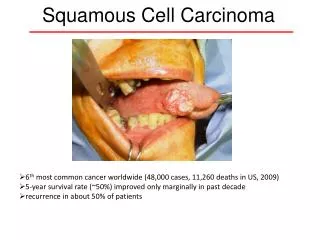
Squamous Cell Carcinoma. Incidence. 2 nd most common skin cancer Behind BCC, accounting for 20% skin cancers Due to propensity to metastasise, makes them responsible for majority of NMSC deaths. Pathogenesis. UV Incidence doubles with 8-10 degrees decrease in latitude

E N D
Incidence • 2nd most common skin cancer • Behind BCC, accounting for 20% skin cancers • Due to propensity to metastasise, makes them responsible for majority of NMSC deaths
Pathogenesis • UV • Incidence doubles with 8-10 degrees decrease in latitude • Induces formation of pyrimidine dimers resulting in DNA point mutations • Causes mutations in p53 tumour suppressor gene
Skin pigmentation • Age • Primary dermatoses – xeroderma pigmentosa, oculocutaneous albinism
Immunosuppression – due to immunosuppressive drugs, UVR, viral infection esp HPV • Reversed ratio of BCC:SCC, SCC being 3x more common in transplant patients • Higher rates – cumulative risk of SCC/ BCC in heart transplant recipient is 3% at one year, 21% at 5 years, 35% at 10 years
Histological subtypes • Pleomorphic • Adenoid/Acantholytic • Simplex • Small cell • Verrucous • Keratoacanthoma • Actinic keratosis • Bowenoid/Erythoplasia of Queyrat
Simplex • Majority of SCCs • Atypical keratinocytes develop within epidermis and invade the dermis • Tumour cells are enlarged, hyperchromatic, variably pleomorphic nuclei, prominent mitotic activity • Keratin pearls
Actinic Keratosis • Also SCC in situ, micro invasive SCC, as there is considerable overlap in the histology • Atypical keratinocytes that have not breached the dermal barrier • SCCIS is typically full thickness keratinocyte atypia
Rate of malignant transformation is 0.1% per lesion per year • About 16% will eventually transform • Can progress to other skin cancers such as sebaceous carcinoma
Pleomorphic • AKA spindle / sarcomatoid, RARE • Associated with previous trauma or RTX • Most commonly found on face or sun exposed areas of elderly • Commonly ulcerate, but may present as an exophytic mass
Microscopically whorls of atypical squamous cells co-mingle with collagen fibres • Pleomorphic giant/spindle cells may be present • Neoplastic keratinocytes have hyperchromatic eosinophilic cytoplasm, elongated, pleomorphic and veiscular nuclei with multiple nucleoli
Small cell • May resemble metastatic small cell neuroendocrine carcinoma or Merkel cell carcinoma • Invades in cohesive nests with adjacent intense inflammatory and desmoplastic host response • Stains for cytokeratin, but may stain for neuron specific enolase (NSE), a neuroendocrine marker
Verrucous • Exophytic or endophytic masses growing at sites of chronic irritation • Slowly locally invasive, little or no propensity to metastasise • Morphologically appear well differentiated with little atypia • Thickened papillae composed with well differentiated squamous cells invading into dermis
Verrucous • 3 distinct clinicopathologic subtypes • Oral • Associated with tobacco chewing, betel nut chewing, HPV, poor oral hygiene • Typically wart like white/gray lesion • Well differentiated • Plantar • Many crypt like openings • Slowly enlarging, fleshy pink exophytic mass • Verrucous hyper/para keratotic component, epithelial crypts with keratinaceous debris • Buschke-Loewenstein • Anogenital type, described by B-L in 1925 • Occur most commonly in uncircumcised men under 50, associated with HPV 6 & 11 • Present as caulflower like lesions most commonly on glans penis • Extensive verrucous acanthosis with dermal extension, keratinocyte atypia minimal, hypergranulosis and crypt/sinus formation
Keratoacanthoma • Period of rapid growth lasts 4-8 weeks • Potential for spontaneous involution usually within 4-6 months, sometimes with considerable scarring • Clinically tend to be rapid growing smooth, firm nodule with central keratin plug
Histologically difficult to distinguish between benign KA and SCC KA type, so being amalgamated by histopathologists • Atypical squamous proliferation with intradermal invasion • Typically crateriform architecture with keratin plug and well developed collarette
Adenoid / Acantholytic • Form a pseudoglandular appearance • Cells arranged in cords and nests with clefts produced by acantholysis of cells leaving spaces that superficially resemble glands
Enlarged free floating dysplastic keratinocytes found in lumina • Clinically appear as ulcer on head & neck of men in 5th to 6th decade • High incidence of recurrence after radiation therapy • Tend to be more locally aggressive but metastasise less
Bowenoid • Considered to be SCC in situ • Most common site is head and neck, followed by limbs and then trunk • Well demarcated, slow growing, erythematous scaly patch, usually small in size
Histologically shows hyperkeratosis, acanthosis, psoriasiform hyperplasia, full thickness atypia, loss of polarity reflecting cessation of maturation • When neoplastic keratinocytes invade the dermis, this lesion is termed Bowenoid SCC • Especially associated with HPV – HPV2 with extragenital lesions, HPV16 with genital lesions
Metastasis • Overall risk is 2 – 6%, not 0.5% • Recurrent SCC has metastatic rate of 30%, and metastatic cases had a survival rate of 1/3 • Metastases tend to be to regional lymph nodes • Most mets (and local recurrences) are found within first 2 years, and 95% within first 5 years
Risk factors for metastasis and recurrence • Recurrence rate doubled and tripled metastatic rate • Size > 2cm • Grade 3 & 4 vs. Grade 1 & 2 tumours • Well differentiated has recurrence 7%, mod well diff 23%, poor diff 28% • Tumour thickness • 3 year recurrence free survival is 98% for <3.5mm, 84% for > 3.5mm (Breslow thickness) • Rapid growth rate
Sun exposed areas tend to metastasise and recur less than mucosal SCC • Scar SCC are very aggressive • Lip and ear SCC have higher metastatic rate than other head and neck sites (16 & 10%) • Probably due to decreased subcutaneous fat • Nose and scalp, anogenital are intermediate risk • Periungal SCC has high recurrence rate but almost never metastasises • Previous treatment – recurrent cancers have a metastatic rate of 25% • Location – ear 45%, lip 32% metastatic rate
Histopathology • Isolated strands, infiltrative pattern, haphazard growth vs. broad pushing borders • Perineural invasion (occurs in 2-14% SCC, most commonly H&N in elderly men) • Has been quoted as local recurrence 47%, regional mets 35%, distant nodes 15%; so post op RTX commonly offered • NO good evidence that any subtype has greater risk recurrence or metastasis
Immunosuppression • Biologically more aggressive, with higher rates of lymph node metastases and deaths secondary to skin cancer
Surgical Management • Tumours < 2cm diameter are successfully excised 95% of the time with a margin of 4mm, 6mm for high risk cases (Brodland & Zitelli) • Tumours > 2cm diameter require margin of 10mm for equivalent local control rates • Moh’s surgery
Other modalities • Dessication and curettage • Lesions less than 2cm diameter have cure rates of 97-98.8% • Cryosurgery • Well localised, superficial lesions on trunk or limbs • 5FU & Imiquimod & Photodynamic therapy • Useful for actinic keratoses
Radiation Therapy • < 2cm tumours have a cure rate of 95% • Adjunctive RTX must be given within 8 weeks for greatest efficiency • (Late) changes include : • atrophy, fibrosis, hypopigmentation, telangiectasia, ulceration • “As late results of RTX can be poor, it is not recommended for patients under 60 yo with uncomplicated primary SCC” • May hasten natural history of KA